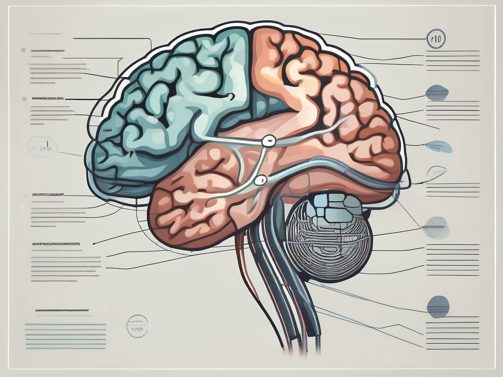
The trochlear nerve, also known as the fourth cranial nerve, is an essential component of the human nervous system. As one of the twelve cranial nerves, it plays a crucial role in controlling various eye movements and allowing us to perceive the world around us. In this article, we will delve into the intricate details of the trochlear nerve, exploring its anatomy, functions, and significance in the field of medical studies.
Understanding the Trochlear Nerve
Before we dive into the specifics of the trochlear nerve, it is important to grasp a basic understanding of this intricate component of our cranial nerves. Situated in the brainstem, this nerve emerges from the dorsal aspect of the midbrain, precisely at the level of the inferior colliculus. Despite its relatively small size, the trochlear nerve has a significant impact on our visual capabilities and overall neurological health.
The trochlear nerve, also known as the fourth cranial nerve, is one of the twelve pairs of cranial nerves that originate from the brain. It is the only cranial nerve that emerges from the dorsal aspect of the brainstem, making it a unique structure in our nervous system. This nerve is responsible for controlling the movement of a specific muscle called the superior oblique muscle, which plays a crucial role in our ability to move our eyes in different directions.
Anatomy of the Trochlear Nerve
Comprised of a singular motor component, the trochlear nerve innervates a specific muscle called the superior oblique muscle. This muscle is primarily responsible for downward eye movement. As the trochlear nerve winds through the subarachnoid space, it undergoes a complex pathway before reaching its target muscle. It even passes through the cavernous sinus, a crucial structure in the skull.
The complex pathway of the trochlear nerve is essential for its proper functioning. It starts at the dorsal aspect of the midbrain, where it emerges and travels through the subarachnoid space, a fluid-filled space surrounding the brain and spinal cord. As it makes its way through this space, it encounters various structures and crosses over the cavernous sinus, a large venous structure located at the base of the skull. This intricate journey ensures that the trochlear nerve reaches its target muscle, the superior oblique muscle, and allows for precise control of eye movements.
Understanding the intricate anatomy of the trochlear nerve allows medical professionals to diagnose any potential issues associated with its function. It is essential to differentiate between trochlear nerve disorders and other cranial nerve abnormalities to ensure accurate diagnosis and effective treatment plans.
Functions of the Trochlear Nerve
The trochlear nerve primarily controls the movement of the superior oblique muscle, allowing us to perform various eye movements. By coordinating the contraction of this muscle, it enables downward, inward, and outward eye movements, contributing to our ability to view objects from different angles and distances.
In addition to its role in eye movements, the trochlear nerve also plays a crucial role in maintaining binocular vision, which allows for optimal depth perception. Binocular vision is the ability to merge the images from both eyes into a single, three-dimensional image. This ability is essential for tasks such as judging distances, catching objects, and navigating our surroundings with precision.
When the trochlear nerve becomes compromised, individuals may experience symptoms such as double vision, eye misalignment, and difficulty moving their eyes in specific directions. These symptoms can significantly impact a person’s quality of life and may require medical intervention to restore proper function of the trochlear nerve.
The Numerical Classification of Cranial Nerves
Within the vast realm of cranial nerves, each nerve possesses a unique numerical classification. The system of numbering these nerves allows medical professionals to navigate the intricate network of structures within the human nervous system.
The cranial nerves, twelve in total, play a crucial role in the functioning of the human body. They are responsible for transmitting sensory information and controlling various motor functions. The numerical classification of these nerves not only provides a means of identification but also serves as a foundation for understanding their anatomical and physiological characteristics.
The Importance of Numbering in Cranial Nerves
Numerical classification eliminates any ambiguity when discussing specific cranial nerves. It provides a clear reference point for medical practitioners, facilitating communication, diagnosis, and treatment. The trochlear nerve, as the fourth cranial nerve, holds a distinct position within this intricate system, allowing for accurate identification and assessment in medical studies and clinical practice.
By assigning a unique number to each cranial nerve, medical professionals can easily refer to a specific nerve during discussions and examinations. This standardized system ensures that there is no confusion or miscommunication when referring to a particular cranial nerve, enabling efficient and effective patient care.
The System Behind the Numerical Classification
The numerical classification of cranial nerves refers to their sequential order from the forebrain to the hindbrain. Rather than chronological order, it follows a systematic arrangement based on their points of origin within the brain. The trochlear nerve, originating from the midbrain, is classified as the fourth nerve. This classification system aids medical professionals in understanding the relationships between cranial nerves and their respective functions.
Understanding the system behind the numerical classification of cranial nerves requires a deep dive into the anatomy of the brain. The cranial nerves emerge from specific regions of the brain, each with its own unique function and distribution. The trochlear nerve, for example, arises from the posterior aspect of the midbrain and innervates the superior oblique muscle of the eye. This specific origin and function contribute to its classification as the fourth cranial nerve.
By organizing the cranial nerves in a numerical sequence, medical professionals can easily identify their locations within the brain and their corresponding functions. This classification system serves as a valuable tool in medical education, allowing students and practitioners to navigate the complex network of cranial nerves with precision and accuracy.
The Trochlear Nerve in Detail
Delving further into the trochlear nerve, we will explore its intricate pathway and the potential disorders associated with its functioning.
The Pathway of the Trochlear Nerve
As mentioned earlier, the trochlear nerve follows a complex pathway before innervating the superior oblique muscle. It wraps around the brainstem and passes through the cavernous sinus, an essential structure housing several vital blood vessels, nerves, and muscles. This intricate route is crucial to ensure the appropriate transmission of signals necessary for eye movement.
The trochlear nerve originates from the dorsal aspect of the midbrain, specifically from the trochlear nucleus. From there, it decussates, meaning it crosses over to the opposite side of the brainstem. This unique feature distinguishes the trochlear nerve from the other cranial nerves, which mostly remain on the same side of the brainstem.
After decussating, the trochlear nerve travels dorsally around the brainstem, wrapping itself around the cerebral peduncle. It then enters the cavernous sinus, a cavity located within the skull, lateral to the sella turcica, which houses the pituitary gland. The cavernous sinus serves as a conduit for multiple structures, including the internal carotid artery, oculomotor nerve, abducens nerve, and of course, the trochlear nerve.
Within the cavernous sinus, the trochlear nerve takes a convoluted path, navigating through the surrounding structures. It carefully weaves its way between the internal carotid artery and the oculomotor nerve, ensuring that it remains protected and undisturbed.
Emerging from the cavernous sinus, the trochlear nerve continues its journey towards the superior oblique muscle. It passes through the superior orbital fissure, a narrow opening located in the sphenoid bone. Finally, it reaches its destination, innervating the superior oblique muscle, which plays a crucial role in eye movement and coordination.
Any disruption in the pathway of the trochlear nerve can lead to debilitating symptoms and hinder an individual’s visual capabilities. Proper understanding of the anatomical route aids medical professionals in diagnosing and treating trochlear nerve disorders effectively.
Disorders Associated with the Trochlear Nerve
Despite its significance in facilitating eye movement, the trochlear nerve can also be susceptible to various disorders. Trochlear nerve palsy, for instance, is a condition characterized by the inability to move the affected eye downward and inward. This condition may result from trauma to the head, infections, autoimmune diseases, or even the presence of tumors.
Other disorders that can affect the trochlear nerve include trochlear nerve neuritis, which is inflammation of the nerve, and trochlear nerve schwannoma, a rare benign tumor that can develop along the nerve.
Diagnosing trochlear nerve disorders requires a comprehensive evaluation by a healthcare professional. The medical history, physical examination, and specialized tests such as magnetic resonance imaging (MRI) or computed tomography (CT) scans may be necessary to assess the condition accurately.
Although this article cannot provide medical advice, it is vital for individuals experiencing any vision-related concerns to consult with a healthcare professional promptly. A thorough evaluation and appropriate diagnostic tests by a qualified medical practitioner are crucial in reaching an accurate diagnosis and establishing an effective treatment plan.
Distinguishing the Trochlear Nerve from Other Cranial Nerves
Understanding the unique characteristics of the trochlear nerve sets it apart from other cranial nerves and aids medical professionals in accurate diagnosis and treatment.
The trochlear nerve, also known as the fourth cranial nerve, plays a crucial role in our visual capabilities. Its distinct features and functions make it essential to differentiate it from other cranial nerves.
Unique Characteristics of the Trochlear Nerve
One distinguishing feature of the trochlear nerve is its long and narrow pathway through the brainstem. Unlike the other cranial nerves emerging directly from the brainstem’s ventral surface, the trochlear nerve emerges from its dorsal aspect, making it anatomically distinct.
As the trochlear nerve emerges from the dorsal aspect of the brainstem, it takes a unique course towards the superior oblique muscle of the eye. This pathway allows it to control the movement of the eye in a specific manner.
Another unique aspect of the trochlear nerve is its decussation or crossing over. Unlike most other cranial nerves, the trochlear nerve partially crosses over to the opposite side of the midbrain, further adding to its distinctive nature.
This crossing over occurs at the level of the superior colliculus, a structure involved in visual processing. The decussation of the trochlear nerve ensures that the appropriate signals are transmitted to the correct eye muscles for coordinated eye movement.
Comparing the Trochlear Nerve with Other Cranial Nerves
Each cranial nerve serves a specific purpose within our nervous system, making it essential to distinguish the trochlear nerve’s characteristics from its counterparts. While some cranial nerves control facial movement or sensory perception, the trochlear nerve’s primary role lies in controlling eye movement, contributing to our visual capabilities.
Unlike the trochlear nerve, which primarily controls eye movement, other cranial nerves have diverse functions. For example, the facial nerve is responsible for facial expression, taste sensation, and tear production. The trigeminal nerve, on the other hand, is involved in facial sensation, chewing, and jaw movement.
Understanding the unique characteristics and functions of each cranial nerve allows medical professionals to accurately diagnose and treat various neurological conditions. By differentiating the trochlear nerve from other cranial nerves, healthcare providers can focus on specific areas of concern related to eye movement and visual coordination.
The Significance of the Trochlear Nerve in Medical Studies
The trochlear nerve holds immense importance in medical studies, aiding researchers and medical professionals in unraveling the complexities of the human nervous system and furthering our understanding of neurological health.
The Role of the Trochlear Nerve in Neurological Health
By thoroughly studying the anatomy and function of the trochlear nerve, researchers can gain insights into normal eye movements and potential abnormalities. This knowledge is paramount in diagnosing various neurologic and ophthalmic disorders.
For instance, understanding how the trochlear nerve controls eye movements can help identify conditions such as trochlear nerve palsy, where the affected individual experiences difficulty moving their eyes vertically. This condition can lead to double vision and challenges in daily activities such as reading or driving.
Moreover, advancements in the field of neurology and neurosurgery rely on a comprehensive understanding of the trochlear nerve. Surgical procedures regarding eye movement disorders often necessitate precise manipulation of this nerve, underscoring its significance in medical practice.
Researchers have also found that the trochlear nerve plays a role in maintaining binocular vision, which is the ability to use both eyes together to perceive depth and accurately judge distances. Understanding the trochlear nerve’s contribution to binocular vision is crucial in diagnosing and treating conditions like strabismus, where the eyes are misaligned and do not work together effectively.
The Trochlear Nerve in Surgical Procedures
Understanding the intricate details of the trochlear nerve assists surgeons in performing delicate procedures, such as trochlear nerve repair or repositioning. These interventions may be necessary in cases of trauma or congenital abnormalities affecting the trochlear nerve’s functioning.
During trochlear nerve repair, surgeons carefully reconnect damaged nerve fibers to restore proper function. This procedure requires a high level of precision and expertise to ensure optimal outcomes for the patient.
Similarly, in cases where the trochlear nerve is positioned incorrectly, surgeons may perform trochlear nerve repositioning surgery. This procedure involves carefully moving the nerve to its correct anatomical position, allowing for improved eye movements and reducing associated symptoms.
It is important to note that any surgical procedure involving the trochlear nerve requires a skilled and experienced surgeon. Consultation with a qualified healthcare professional is crucial to assess individual circumstances and determine the appropriateness of any surgical intervention.
In conclusion, the trochlear nerve plays an integral role in controlling eye movements and maintaining binocular vision. By understanding its anatomy, functions, and unique characteristics, medical professionals can accurately diagnose and treat trochlear nerve disorders. Furthermore, its significance in medical studies and surgical procedures highlights the necessity of thorough research and expertise in the field. Should individuals experience any vision-related concerns or suspect trochlear nerve abnormalities, it is imperative they consult with a healthcare professional for proper evaluation and guidance.
