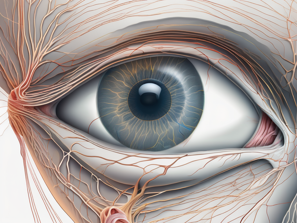
Cranial nerve IV, also known as the trochlear nerve, plays a crucial role in eye movement. Understanding the functions and anatomy of this nerve is essential in comprehending the complexities of its innervation and the impact it has on vision. In this article, we will delve into the intricate details of cranial nerve IV, exploring its function, associated disorders, and its significance in facilitating binocular vision.
Understanding the Function of Cranial Nerve IV
Cranial nerve IV, one of the twelve cranial nerves originating from the brainstem, is unique in its distribution as it is the only nerve to have a contralateral innervation pattern. Despite being the smallest cranial nerve, its role is indispensable in eye movement. The primary function of the trochlear nerve is to innervate the superior oblique muscle, which plays a critical role in eye rotation and stabilization.
The Role of the Trochlear Nerve in Eye Movement
Eye movement is a complex process involving the coordinated contraction and relaxation of multiple muscles. The trochlear nerve serves as the main driver for the superior oblique muscle, enabling downward and inward rotation of the eye. This specific movement aids in maintaining proper eye alignment and contributes to the binocular vision required for depth perception.
In addition to its role in eye movement, the trochlear nerve also plays a crucial role in proprioception. Proprioception refers to the body’s ability to sense the position and movement of its parts. The trochlear nerve provides sensory feedback to the brain about the position and tension of the superior oblique muscle, allowing for fine-tuning of eye movements and coordination.
Furthermore, the trochlear nerve is involved in the regulation of the pupillary light reflex. This reflex is responsible for controlling the size of the pupil in response to changes in light intensity. The trochlear nerve carries signals from the brain to the muscles that control the size of the pupil, ensuring that the appropriate amount of light enters the eye for optimal vision.
Anatomy of the Trochlear Nerve
The trochlear nerve emerges from the midbrain, precisely the dorsal aspect of the brainstem. It possesses the longest intracranial course among all cranial nerves, winding around the inferior colliculus and crossing the midline before reaching its destination. Upon leaving the brainstem, the trochlear nerve follows a complex pathway, passing near the cavernous sinus and superior orbital fissure before finally reaching the superior oblique muscle.
Along its pathway, the trochlear nerve is vulnerable to various pathological conditions that can affect its function. Lesions or damage to the nerve can result in trochlear nerve palsy, a condition characterized by weakness or paralysis of the superior oblique muscle. This can lead to double vision, particularly when looking downward or inward.
In summary, the trochlear nerve is a vital component of the intricate network responsible for eye movement and coordination. Its unique contralateral innervation pattern and role in innervating the superior oblique muscle make it an essential player in maintaining proper eye alignment, depth perception, and pupillary light reflex. Understanding the function and anatomy of the trochlear nerve is crucial for diagnosing and treating conditions that may affect its normal functioning.
The Eye Muscle Innervated by the Trochlear Nerve
The superior oblique muscle, innervated by the trochlear nerve, is a pivotal player in eye movement. It originates from the common tendinous ring, also known as the annulus of Zinn, and passes through a unique structure called the trochlea. The trochlea acts as a pulley, redirecting the muscle’s pull to achieve the desired downward and inward eye rotation.
The trochlear nerve, also known as the fourth cranial nerve, is one of the twelve cranial nerves that emerge directly from the brain. It is the smallest cranial nerve in terms of the number of axons it contains. Despite its small size, the trochlear nerve plays a crucial role in the complex coordination of eye movements.
The Superior Oblique Muscle: An Overview
Located in the upper part of the eye socket, the superior oblique muscle has a fascinating structure. It forms a tendon that attaches to the eyeball near the insertion site of another significant eye muscle, the superior rectus muscle. This anatomical arrangement enables the trochlear nerve to exert its control over the superior oblique muscle, facilitating precise eye movement.
The superior oblique muscle is responsible for a variety of eye movements. It helps rotate the eye downward and inward, allowing us to focus on objects located below our line of sight. Additionally, it assists in the torsional movement of the eye, which is the rotation of the eyeball around its vertical axis. This rotational movement is essential for maintaining proper alignment and coordination between both eyes.
How the Trochlear Nerve Controls the Superior Oblique Muscle
The trochlear nerve employs a unique mechanism to control the superior oblique muscle. By sending signals to the muscle fibers, it initiates the contraction responsible for eye rotation. This intricate coordination allows for precise control and adjustments of eye movement, ultimately contributing to vision stabilization and the ability to focus on objects of interest.
The trochlear nerve’s control over the superior oblique muscle is finely tuned to ensure smooth and coordinated eye movements. Dysfunction or damage to the trochlear nerve can result in a condition called trochlear nerve palsy, which can lead to various eye movement abnormalities. These abnormalities may include vertical double vision, difficulty looking downward, and a head tilt to compensate for the impaired eye movement.
In summary, the superior oblique muscle, innervated by the trochlear nerve, is a remarkable structure that plays a vital role in eye movement. Its unique anatomical arrangement and precise control mechanisms allow for precise and coordinated eye movements, contributing to our ability to perceive the world around us.
Disorders Related to the Trochlear Nerve
Despite its small size, the trochlear nerve can be subjected to various disorders that impede its proper functioning. These conditions can lead to noticeable symptoms, necessitating diagnosis and treatment by medical professionals. If you experience any eye movement abnormalities or related issues, it is vital to consult with a doctor for appropriate evaluation and guidance.
The trochlear nerve, also known as the fourth cranial nerve, plays a crucial role in controlling the movement of the superior oblique muscle, which is responsible for rotating the eye downward and inward. Any disruption or damage to this nerve can result in significant visual disturbances and impairments.
One of the most common symptoms of trochlear nerve damage is double vision, also known as diplopia. This occurs when the eyes are unable to align properly, causing two separate images to be perceived instead of one. The misalignment can be horizontal, vertical, or even diagonal, depending on the extent of the nerve damage.
In addition to double vision, individuals with trochlear nerve disorders may experience a feeling that one eye is higher or lower than the other. This sensation, known as ocular tilt reaction, can be disorienting and affect depth perception. It can make everyday tasks such as reading or navigating obstacles challenging and frustrating.
Difficulties with downward and inward eye movements are also common in trochlear nerve disorders. These impairments can make it difficult to read, as the eyes may not be able to move smoothly across the lines of text. Furthermore, navigating obstacles or stairs can become hazardous, as the eyes may not be able to coordinate properly to assess depth and distance accurately.
Symptoms of Trochlear Nerve Damage
Trochlear nerve damage can manifest through a range of symptoms, including double vision (also known as diplopia), eye misalignment, and feeling that one eye is higher or lower than the other. Difficulties with downward and inward eye movements may also be present, impairing the ability to read or navigate obstacles correctly. If any of these symptoms occur, prompt medical attention is advised to determine the underlying cause and appropriate course of action.
It is important to note that trochlear nerve disorders can occur due to various factors, including trauma, tumors, infections, or even congenital abnormalities. Understanding the underlying cause is crucial in determining the most effective treatment approach.
Diagnosis and Treatment of Trochlear Nerve Disorders
Diagnosing trochlear nerve disorders requires a comprehensive evaluation by a healthcare professional, often involving a detailed medical history, physical examination, and potentially imaging studies. The doctor will assess the patient’s symptoms, perform a thorough eye examination, and may order additional tests such as magnetic resonance imaging (MRI) or computed tomography (CT) scans to visualize the nerve and surrounding structures.
Once a diagnosis is made, the treatment plan will depend on the specific disorder and the severity of the symptoms. In some cases, non-invasive approaches such as eye exercises or prism glasses may be recommended to help correct the misalignment and improve eye movements. These exercises aim to strengthen the eye muscles and improve coordination.
However, in more severe cases or when conservative measures fail to provide relief, surgical intervention may be necessary. Surgical options can include procedures to correct muscle imbalances or reposition the eye to improve alignment. The specific surgical technique will depend on the individual’s condition and the underlying cause of the trochlear nerve disorder.
It is essential for individuals with trochlear nerve disorders to consult with a doctor who specializes in ophthalmology or neurology. These specialists have the expertise and knowledge to provide tailored insights and guidance based on each individual’s unique circumstances. Early diagnosis and appropriate treatment can significantly improve the prognosis and quality of life for individuals with trochlear nerve disorders.
The Impact of Trochlear Nerve Function on Vision
Understanding the crucial role of the trochlear nerve in facilitating precise eye movement sheds light on its significant impact on vision. The ability to coordinate both eyes, align their movements, and maintain binocular vision is essential for daily activities such as reading, driving, and depth perception.
When we think about vision, we often focus on the eyes themselves. However, the complex network of nerves that control eye movement plays a vital role in how we see the world around us. One of these important nerves is the trochlear nerve, also known as cranial nerve IV.
The Role of the Trochlear Nerve in Binocular Vision
Binocular vision, the simultaneous use of both eyes to perceive a single, integrated image, allows for enhanced depth perception and improved visual acuity. The trochlear nerve, through its innervation of the superior oblique muscle, ensures that each eye is properly aligned and contributes to the overall visual experience.
Imagine trying to catch a ball without the ability to accurately judge its distance or trajectory. This is where the trochlear nerve steps in, helping us to precisely track moving objects and maintain a clear and focused image. Without the trochlear nerve’s influence, our ability to perceive depth and accurately judge distances would be severely compromised.
Vision Problems Associated with Trochlear Nerve Damage
Trochlear nerve damage can disrupt the delicate balance necessary for binocular vision, leading to vision problems. These problems may include blurred or double vision, difficulties with depth perception, and challenges in accurately judging distances.
Imagine waking up one day and finding that your vision has suddenly become blurry or that you see two of everything. These are some of the distressing symptoms that can occur when the trochlear nerve is damaged. Simple tasks like reading a book or driving a car become incredibly challenging and frustrating.
Early detection and appropriate treatment are crucial in mitigating the impact of trochlear nerve damage on vision. If you notice any changes in your vision or experience difficulties with eye movement, it is important to seek medical attention promptly. An eye care professional can conduct a thorough examination and determine the underlying cause of your symptoms.
In conclusion, cranial nerve IV, the trochlear nerve, holds a vital position in the intricate network of eye movement control. From its innervation of the superior oblique muscle to its contributions to binocular vision, understanding the functions, anatomy, and associated disorders of this nerve provides valuable insights into the complexities of vision.
Our eyes are truly remarkable organs, allowing us to experience the world in vibrant detail. However, it is important to remember that vision is not solely dependent on the eyes themselves. The intricate interplay between nerves, muscles, and the brain is what enables us to see and interpret the world around us.
If you experience any eye movement abnormalities or suspect trochlear nerve-related issues, it is essential to consult with a healthcare professional to ensure appropriate assessment and guidance tailored to your unique circumstances. Taking care of your vision is taking care of your overall well-being.
