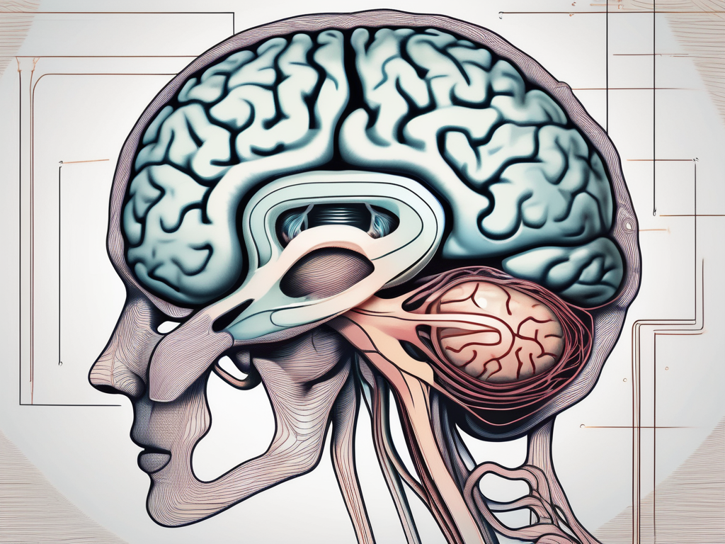
The trochlear nerve plays a crucial role in our ability to move our eyes and control their position. It is responsible for coordinating the movement of the superior oblique muscle, which helps us move our eyes downward and inward. However, like any other nerve in our body, the trochlear nerve can be affected by various conditions and injuries.
Understanding the Trochlear Nerve
The trochlear nerve, also known as cranial nerve IV, is the smallest of the twelve cranial nerves in our body. It plays a crucial role in our visual system, specifically in controlling the movement of the eye. Let’s dive deeper into the fascinating world of the trochlear nerve and explore its anatomy and function.
Anatomy of the Trochlear Nerve
The trochlear nerve emerges from the posterior aspect of the midbrain, near the brainstem’s superior colliculus. It is the only cranial nerve that exits the brainstem dorsally, meaning it emerges from the back of the brainstem. This unique anatomical feature sets it apart from the other cranial nerves.
From its origin, the trochlear nerve follows a complex path, looping around the brainstem to reach the superior oblique muscle. This intricate route ensures that the nerve fibers reach their target muscle precisely, allowing for coordinated eye movements. The trochlear nerve’s journey resembles a carefully orchestrated dance, with each step serving a specific purpose.
As the trochlear nerve courses around the brainstem, it passes through the cavernous sinus, a cavity located on either side of the sella turcica (a bony structure in the skull). This proximity to the cavernous sinus exposes the trochlear nerve to potential compression or damage in certain medical conditions, leading to trochlear nerve palsy.
Function of the Trochlear Nerve
The primary function of the trochlear nerve is to control the contraction of the superior oblique muscle, one of the six extraocular muscles responsible for eye movements. When the trochlear nerve fires, it causes the superior oblique muscle to contract, resulting in a downward and inward movement of the eye.
This downward and inward eye movement is crucial for various visual tasks. When we read, the trochlear nerve helps us smoothly track the lines of text, ensuring that our eyes move in a coordinated and controlled manner. Similarly, when we focus on near objects, such as when working on a computer or reading a book, the trochlear nerve plays a vital role in adjusting our eye position to maintain clear vision.
Moreover, the trochlear nerve contributes to the ability to track moving objects across our field of vision. Whether it’s following a flying bird or catching a ball, the trochlear nerve ensures that our eyes can accurately track the object’s trajectory, allowing us to perceive depth and movement.
Given its role in controlling eye movements, any dysfunction or damage to the trochlear nerve can lead to various visual disturbances. Trochlear nerve palsy, characterized by weakness or paralysis of the superior oblique muscle, can result in double vision, difficulty looking downward, and an abnormal head tilt to compensate for the eye misalignment.
In conclusion, the trochlear nerve is a remarkable cranial nerve that orchestrates the precise movements of the superior oblique muscle. Its intricate anatomy and crucial function in eye movements make it a fascinating subject of study in the field of neuroanatomy and ophthalmology.
Symptoms of Trochlear Nerve Damage
Damage to the trochlear nerve can result in a variety of symptoms, which may vary depending on the extent and location of the damage. It is important to note that these symptoms can also be indicative of other underlying conditions, so a proper medical evaluation is necessary.
Vision Problems
Individuals with trochlear nerve damage may experience a range of vision problems. These can include double vision, difficulty in moving the affected eye downward or inward, and decreased coordination between both eyes.
Double vision, also known as diplopia, occurs when the brain receives conflicting signals from the affected eye and the healthy eye. This can make it challenging for individuals to focus on objects, read, or perform daily activities that require clear vision. The misalignment of the eyes can cause objects to appear blurry or distorted, making it difficult to navigate the world around them.
In addition to double vision, trochlear nerve damage can also lead to difficulty in moving the affected eye downward or inward. This can result in limited eye movement and may affect an individual’s ability to track objects or shift their gaze. Tasks that require looking down, such as reading or walking down stairs, can become particularly challenging.
Furthermore, trochlear nerve damage can cause decreased coordination between both eyes. This can lead to a condition known as strabismus, where the eyes are misaligned and point in different directions. Strabismus can affect depth perception and make it difficult to judge distances accurately. It can also impact an individual’s self-esteem and social interactions, as the misalignment of the eyes may be noticeable to others.
Head and Eye Pain
Trochlear nerve damage can also be associated with head and eye pain. This can be a result of strain on the extraocular muscles or the presence of associated conditions.
The extraocular muscles, responsible for controlling eye movements, can become strained when the trochlear nerve is damaged. This strain can lead to discomfort and pain in the eye area, which may radiate to the surrounding regions of the head. The pain can range from mild to severe and may worsen with eye movement or prolonged use of the affected eye.
In some cases, trochlear nerve damage can be accompanied by associated conditions such as migraines or cluster headaches. These types of headaches can cause intense pain, often described as a throbbing or pulsating sensation, and may be accompanied by other symptoms such as nausea, sensitivity to light, and sound. The presence of these associated conditions can further contribute to head and eye pain in individuals with trochlear nerve damage.
Diagnostic Procedures for Trochlear Nerve Disorders
When symptoms suggestive of trochlear nerve damage are present, diagnostic evaluation is essential to determine the underlying cause. A thorough examination may involve:
Physical Examination
A physical examination allows a healthcare professional to assess the range of motion of the affected eye and evaluate any observable abnormalities. Various eye movement tests may be performed to assess the coordination and function of the superior oblique muscle.
During the physical examination, the healthcare professional will carefully observe the patient’s eye movements, looking for any signs of weakness or limited range of motion. They may ask the patient to follow their finger or a moving object with their eyes, checking for any abnormalities in the smoothness or accuracy of the eye movements.
In addition to assessing the eye movements, the healthcare professional may also perform other tests to evaluate the function of the trochlear nerve. These tests may include checking the patient’s ability to focus on objects at different distances, assessing their depth perception, and evaluating their ability to maintain eye alignment.
Imaging Techniques
In some cases, imaging techniques such as MRI (Magnetic Resonance Imaging) or CT (Computed Tomography) scans may be used to visualize the brainstem and surrounding structures. These imaging modalities can provide detailed information about potential causes of trochlear nerve damage, such as tumors, inflammation, or other structural abnormalities.
MRI scans use powerful magnets and radio waves to create detailed images of the brain and surrounding tissues. This imaging technique can help identify any abnormalities or lesions that may be affecting the trochlear nerve. CT scans, on the other hand, use X-rays to create cross-sectional images of the body. These scans can provide valuable information about the bony structures and can help identify any fractures or other injuries that may be contributing to the trochlear nerve disorder.
During the imaging procedure, the patient will lie on a table that slides into the scanner. They may need to remain still for a certain period of time while the images are being taken. The healthcare professional may also use contrast agents, such as a dye injected into the bloodstream, to enhance the visibility of certain structures or abnormalities.
Once the imaging is complete, the healthcare professional will carefully analyze the images to identify any potential causes of the trochlear nerve disorder. They will look for any signs of tumors, inflammation, or other structural abnormalities that may be affecting the nerve’s function.
Interpreting Test Results
Interpreting the test results correctly is crucial in understanding the trochlear nerve’s condition and determining appropriate treatment strategies. It requires expertise and a comprehensive understanding of the nervous system and associated disorders.
The trochlear nerve, also known as cranial nerve IV, plays a vital role in eye movement. It is responsible for the movement of the superior oblique muscle, which helps rotate the eye downward and outward. When the trochlear nerve is damaged or dysfunctional, it can lead to various visual impairments and difficulties in eye coordination.
Normal vs Abnormal Findings
Normal test findings suggest that the trochlear nerve and associated structures are functioning properly. The eye movements are coordinated, and there are no signs of weakness or limitations. However, abnormal findings can indicate various conditions, including nerve trauma, tumors, congenital abnormalities, or other neurological disorders.
One common abnormal finding is trochlear nerve palsy, which refers to the paralysis or weakness of the trochlear nerve. This condition can result from head trauma, infections, or even certain medications. Trochlear nerve palsy often leads to double vision, particularly when looking downward or to the side. It can significantly impact a person’s daily activities and overall quality of life.
Potential Disorders and Conditions
There are several conditions that can result in trochlear nerve damage or dysfunction. These range from congenital abnormalities, such as trochlear nerve palsy, to acquired conditions like trauma, infections, or inflammatory diseases.
In some cases, trochlear nerve damage can occur during birth due to factors like a difficult delivery or abnormal positioning of the baby’s head. This congenital condition may require early intervention and specialized treatment to improve eye coordination and prevent long-term visual impairments.
Trauma, such as a head injury or facial fracture, can also lead to trochlear nerve damage. The forceful impact can disrupt the nerve’s normal function, resulting in eye movement difficulties. Prompt medical attention and appropriate rehabilitation are essential in these cases to optimize recovery and minimize long-term complications.
Infections, such as meningitis or encephalitis, can also affect the trochlear nerve. These inflammatory conditions can cause nerve inflammation and damage, leading to visual disturbances and eye movement abnormalities. Timely diagnosis and targeted treatment are crucial in managing these infections and preventing further nerve damage.
Furthermore, certain autoimmune diseases, such as multiple sclerosis, can also impact the trochlear nerve. In these cases, the immune system mistakenly attacks the protective covering of the nerve, disrupting its normal function. Managing the underlying autoimmune condition is vital to prevent further nerve damage and preserve visual function.
A thorough analysis of the test results helps healthcare professionals identify the specific disorder or condition causing trochlear nerve damage. This information guides the development of an individualized treatment plan, which may include medication, physical therapy, or even surgical interventions.
Treatment Options for Trochlear Nerve Damage
Once a diagnosis is made, the treatment approach for trochlear nerve damage depends on the underlying cause and the severity of the symptoms. It is important to consult with a healthcare professional for individualized management strategies.
Trochlear nerve damage can be a challenging condition to manage, but there are various treatment options available to help alleviate symptoms and promote recovery. Non-surgical interventions and surgical procedures are two main approaches that healthcare professionals may consider.
Non-Surgical Interventions
Non-surgical interventions are often the first line of treatment for trochlear nerve damage. These interventions focus on managing symptoms and promoting the nerve’s recovery. One of the key aspects of non-surgical interventions is pain management. Healthcare professionals may prescribe pain medications or recommend non-pharmacological approaches such as heat or cold therapy to help alleviate discomfort.
In addition to pain management, eye exercises play a crucial role in the non-surgical treatment of trochlear nerve damage. These exercises aim to improve eye coordination and strengthen the affected eye muscles. By engaging in specific eye movements and visual tasks, patients can gradually regain control over their eye movements and reduce symptoms such as double vision or eye misalignment.
Protective eyewear is another important aspect of non-surgical interventions. It helps prevent further damage to the trochlear nerve and provides support to the affected eye. Healthcare professionals may recommend specialized eyewear or suggest modifications to existing eyeglasses to ensure optimal protection and comfort.
Addressing underlying conditions that contribute to trochlear nerve damage is also a crucial part of non-surgical interventions. For example, if the nerve damage is caused by an underlying medical condition such as diabetes or hypertension, healthcare professionals will focus on managing and treating these conditions to prevent further nerve damage and promote overall healing.
Surgical Procedures
In some cases, surgical intervention may be necessary to correct or alleviate the underlying cause of trochlear nerve damage. These procedures aim to repair damaged structures or relieve pressure on the nerve, restoring its normal function.
One surgical option for trochlear nerve damage is decompression surgery. This procedure involves removing any structures or tissues that may be compressing or impinging on the nerve. By relieving the pressure, the nerve can regain its normal function and symptoms can be alleviated.
In cases where the trochlear nerve is severely damaged or completely severed, nerve repair surgery may be considered. This procedure involves reconnecting the damaged nerve ends or using a nerve graft to bridge the gap between the severed ends. Nerve repair surgery aims to restore the continuity of the nerve and promote functional recovery.
Another surgical option for trochlear nerve damage is muscle surgery. This procedure involves repositioning or adjusting the eye muscles to improve eye alignment and reduce symptoms such as double vision. By modifying the positioning of the muscles, the affected eye can regain its normal movement and coordination.
It is important to note that the decision to undergo surgical intervention for trochlear nerve damage is based on individual circumstances and the recommendations of healthcare professionals. They will carefully assess the severity of the nerve damage, the potential benefits of surgery, and any associated risks before recommending surgical intervention.
In conclusion, the treatment options for trochlear nerve damage are diverse and tailored to individual needs. Non-surgical interventions focus on symptom management, eye exercises, protective eyewear, and addressing underlying conditions. Surgical procedures aim to correct or alleviate the underlying cause of the nerve damage, restoring its normal function. Consulting with a healthcare professional is essential to determine the most suitable treatment approach for trochlear nerve damage.
Recovery and Rehabilitation from Trochlear Nerve Damage
Recovery from trochlear nerve damage can vary depending on the cause, extent of nerve involvement, and individual factors. A comprehensive rehabilitation plan is crucial to optimize recovery and regain normal eye movements.
Physical Therapy and Exercises
Physical therapy plays a vital role in rehabilitating the trochlear nerve. It may involve specific exercises to strengthen and retrain the eye muscles, enhance coordination between both eyes, and improve overall eye movement control.
Coping with Long-Term Effects
In cases where trochlear nerve damage results in long-term effects, it is essential to develop coping strategies to manage any residual symptoms. This may involve lifestyle modifications, assistive devices, and ongoing support from healthcare professionals.
In conclusion, testing for trochlear nerve damage requires a careful evaluation of symptoms, thorough physical examination, and appropriate diagnostic procedures. Since the causes and severity of trochlear nerve disorders can vary, consultation with a healthcare professional is strongly recommended for a comprehensive evaluation and tailored treatment plan.
