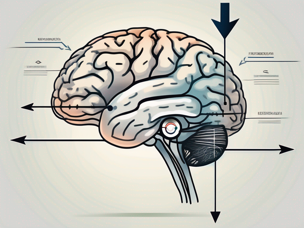
The trochlear nerve, also known as cranial nerve IV, plays a crucial role in the complex process of eye movement. Specifically, it is responsible for downward movement of the eye. Understanding the anatomy and function of this nerve is essential to comprehending how it achieves this intricate task.
Understanding the Trochlear Nerve
The trochlear nerve, also known as the fourth cranial nerve, is a fascinating component of the human nervous system. It holds the distinction of being the smallest of the twelve cranial nerves and originates from the dorsal aspect of the brainstem. Its intricate structure and function play a crucial role in eye movement and coordination.
The trochlear nerve contains motor fibers that emerge from the trochlear nucleus, a region located in the midbrain. These fibers extend to the superior oblique muscle of the eye, which is responsible for various eye movements, including a downward rotation. This unique positioning allows the trochlear nerve to influence the downward movement of the eye, contributing to our ability to perceive the world around us.
Anatomy of the Trochlear Nerve
The trochlear nerve follows a distinct pathway within the brain, showcasing the intricacy of its anatomical course. It traverses the midbrain, a vital region that serves as a relay station for sensory and motor signals. As the trochlear nerve continues its journey, it crosses the cavernous sinus, a venous structure located within the skull. This crossing further highlights the complex nature of its route.
After its passage through the cavernous sinus, the trochlear nerve enters the orbit through the superior orbital fissure, a narrow opening in the bony structure of the skull. Once inside the orbit, it innervates the superior oblique muscle, establishing a direct connection between the nerve and the muscle responsible for downward eye movement.
The superior oblique muscle, one of the six extraocular muscles, plays a crucial role in eye movement coordination. Its unique arrangement and connection to the trochlear nerve ensure that it receives the necessary signals to execute a downward rotation of the eye, contributing to our ability to explore our visual environment.
Function of the Trochlear Nerve
The primary function of the trochlear nerve is to provide motor innervation to the superior oblique muscle. This muscle’s role in eye movement coordination cannot be overstated, as it facilitates the downward rotation of the eye.
When the brain signals the trochlear nerve to initiate a downward eye movement, the nerve transmits the appropriate electrical impulses to the superior oblique muscle. These impulses serve as a command for the muscle to contract, resulting in a downward rotation of the eye. This intricate interplay between the trochlear nerve and the superior oblique muscle allows us to navigate our visual surroundings with precision and accuracy.
In conclusion, the trochlear nerve, despite being the smallest of the twelve cranial nerves, plays a crucial role in eye movement coordination. Its unique anatomical course and connection to the superior oblique muscle allow it to influence the downward rotation of the eye, contributing to our ability to perceive and interact with the world around us.
The Role of the Trochlear Nerve in Eye Movement
The mechanism behind the trochlear nerve’s involvement in eye movement is fascinating. Not only does it ensure smooth and coordinated downward motion, but it also interacts synergistically with other eye muscles to maintain visual stability.
The trochlear nerve, also known as cranial nerve IV, is responsible for innervating the superior oblique muscle. This muscle plays a crucial role in the downward movement of the eye. Without the trochlear nerve’s stimulation, the superior oblique muscle would not be able to function effectively, leading to impaired downward eye movement.
Mechanism of Downward Eye Movement
During a downward eye movement, the trochlear nerve’s stimulation of the superior oblique muscle works in harmony with the actions of the superior rectus and inferior rectus muscles. While the superior oblique muscle depresses the eye, the superior and inferior rectus muscles counteract this movement, ensuring precise control and alignment.
The superior rectus muscle primarily elevates the eye, allowing us to look upwards. On the other hand, the inferior rectus muscle primarily depresses the eye, enabling us to look downwards. The coordinated action of these muscles, guided by the trochlear nerve, ensures smooth and accurate downward eye movement.
This intricate interplay between these muscles maintains balanced eye movement, allowing us to comfortably track objects or navigate our surroundings without visual disturbances.
Interaction with Other Eye Muscles
The trochlear nerve’s communication with other eye muscles extends beyond the superior oblique. For instance, it interacts with the oculomotor nerve, also known as cranial nerve III, which controls several other extraocular muscles. This intricate network synergizes their movements, optimizing visual functionality.
The oculomotor nerve innervates the superior rectus, inferior rectus, and inferior oblique muscles, among others. These muscles work in coordination with the superior oblique muscle innervated by the trochlear nerve to enable various eye movements, including vertical and rotational motions.
Through these complex interactions, the trochlear nerve contributes to the intricate ballet of eye movements, ensuring accurate downward rotation while preserving overall visual balance.
Understanding the role of the trochlear nerve in eye movement provides insight into the remarkable complexity and precision of our visual system. The ability to move our eyes with such control and coordination is a testament to the intricate interplay between various cranial nerves and extraocular muscles.
Further research into the trochlear nerve’s function and its interactions with other components of the visual system may uncover even more fascinating details about the mechanisms behind eye movement.
Disorders Related to the Trochlear Nerve
The trochlear nerve, also known as cranial nerve IV, plays a crucial role in eye movement and control. It is the smallest of the twelve cranial nerves and innervates the superior oblique muscle, which is responsible for rotating the eye downward and inward. Though the trochlear nerve operates with remarkable precision, its dysfunction can result in various visual disturbances that can significantly impact a person’s quality of life.
When the trochlear nerve experiences dysfunction, it can lead to a condition known as trochlear nerve palsy. Trochlear nerve palsy is characterized by the impairment of the superior oblique muscle, which affects the eye’s ability to rotate efficiently. This impairment often manifests in symptoms such as double vision, difficulty focusing, or a feeling of imbalance. These symptoms are most prominent during downward eye movements, as the affected muscle struggles to perform its intended function.
If you are experiencing any of these symptoms, it is essential to consult with an ophthalmologist or neurologist who can perform a thorough evaluation and provide an accurate diagnosis. The diagnosis of trochlear nerve dysfunction requires a comprehensive assessment involving a detailed medical history, physical examination, and potentially specialized tests, such as neuroimaging.
During the evaluation, the healthcare professional will carefully review your medical history to identify any potential risk factors or underlying conditions that may contribute to the trochlear nerve dysfunction. They will also conduct a physical examination, which may involve assessing eye movements, checking for muscle weakness or imbalances, and evaluating overall visual function.
In some cases, additional tests may be necessary to confirm the diagnosis. Neuroimaging, such as magnetic resonance imaging (MRI) or computed tomography (CT) scans, can help identify any structural abnormalities or nerve compression that may be causing the trochlear nerve dysfunction. These tests provide valuable insights into the underlying cause, which can range from nerve compression due to tumors or vascular malformations to traumatic injury or inflammation.
Once a diagnosis is confirmed, the healthcare professional will discuss appropriate treatment options based on the specific etiology of the trochlear nerve dysfunction. Treatment may involve medication to manage symptoms, vision therapy to improve eye coordination and control, or, in severe cases, surgery to correct structural abnormalities or release nerve compression.
It is important to note that each case of trochlear nerve dysfunction is unique, and treatment plans should be tailored to individual needs. Only a trained healthcare professional can suggest appropriate treatment after a thorough evaluation. By seeking timely medical attention and following the recommended treatment plan, individuals with trochlear nerve dysfunction can improve their visual function and enhance their overall quality of life.
The Impact of Trochlear Nerve on Vision
A healthy trochlear nerve plays a critical role in maintaining optimal vision. When this nerve functions properly, its coordinated communication with various eye muscles ensures that our eyes move fluidly and accurately.
The trochlear nerve, also known as the fourth cranial nerve, originates in the brainstem and controls the superior oblique muscle of the eye. This muscle is responsible for downward and inward eye movements, allowing us to look down and towards the nose. The trochlear nerve’s precise control over this muscle is essential for coordinated eye movements and binocular vision.
When trochlear nerve dysfunction occurs, it can result in vision changes such as double vision, reduced visual acuity, or difficulty tracking moving objects. These visual disturbances can disrupt daily activities and impact overall visual perception.
Individuals with trochlear nerve dysfunction may experience diplopia, commonly known as double vision. This occurs when the eyes are unable to align properly, causing two images to be perceived instead of one. Double vision can make it challenging to read, drive, or perform tasks that require visual accuracy.
Reduced visual acuity is another common symptom of trochlear nerve dysfunction. It can manifest as blurred vision or the inability to see objects clearly. This can significantly impact one’s ability to navigate the environment and perform daily tasks with ease.
Difficulty tracking moving objects is yet another vision change associated with trochlear nerve dysfunction. When the trochlear nerve is not functioning correctly, the eyes may struggle to smoothly follow objects in motion. This can make activities like playing sports or watching fast-paced movies challenging and frustrating.
If you are experiencing vision changes or suspect trochlear nerve involvement, it is vital to seek professional medical advice promptly. An eye care specialist can thoroughly examine your visual system and provide appropriate guidance based on their clinical expertise.
Preventing Vision Problems Related to Trochlear Nerve Dysfunction
Prevention of vision problems related to trochlear nerve dysfunction begins with maintaining an overall healthy lifestyle. Regular exercise, a well-balanced diet, and maintaining good eye health habits can significantly contribute to preserving optimal visual function.
Regular exercise promotes blood circulation and oxygenation, which is crucial for maintaining the health of the nerves and muscles involved in vision. Engaging in activities such as walking, jogging, or yoga can help keep your eyes and the trochlear nerve in good shape.
A well-balanced diet rich in essential nutrients, vitamins, and minerals is also essential for maintaining optimal visual function. Foods like leafy greens, carrots, citrus fruits, and fish contain nutrients that support eye health and may help prevent nerve dysfunction.
Maintaining good eye health habits, such as wearing protective eyewear in hazardous environments and taking regular breaks from screen time, can also contribute to preventing vision problems related to trochlear nerve dysfunction.
Should you have any concerns about your vision, it is vital to prioritize regular eye exams. These routine evaluations enable early detection of any potential issues and allow your eye care professional to take appropriate measures to address and manage them effectively.
The Future of Trochlear Nerve Research
As our understanding of the trochlear nerve and its role in eye movement continues to advance, ongoing research explores promising avenues to improve diagnosis, treatment, and prevention of trochlear nerve-related disorders.
The trochlear nerve, also known as the fourth cranial nerve, plays a crucial role in eye movement. It innervates the superior oblique muscle, which is responsible for downward eye movement and inward rotation of the eye. Dysfunction of the trochlear nerve can lead to a range of visual disturbances, including double vision, difficulty focusing, and impaired depth perception.
Current Research Trends
Current research is primarily focused on refining diagnostic methods and enhancing our understanding of the precise mechanisms underlying trochlear nerve dysfunction. Scientists and researchers are investigating potential therapeutic interventions to mitigate the impact of trochlear nerve-related disorders on visual function and overall quality of life.
One area of research interest is the development of advanced imaging techniques to visualize the trochlear nerve and its surrounding structures with greater precision. High-resolution magnetic resonance imaging (MRI) and diffusion tensor imaging (DTI) are being used to map the course of the trochlear nerve and identify any abnormalities or damage. These imaging modalities provide valuable insights into the structural integrity of the nerve and aid in accurate diagnosis.
Another research focus is the exploration of genetic factors that may contribute to trochlear nerve dysfunction. Genetic studies are being conducted to identify specific gene mutations or variations that could increase the risk of developing trochlear nerve-related disorders. Understanding the genetic basis of these conditions may pave the way for targeted therapies and personalized treatment approaches.
Potential Breakthroughs in Understanding and Treatment
Advancements in technology and medical imaging techniques provide hope for improved diagnosis and treatment options for trochlear nerve-related disorders. Additionally, ongoing studies shed light on potential breakthroughs in rehabilitation and vision therapy, which may enhance the management of these conditions.
One exciting area of research is the use of virtual reality (VR) and augmented reality (AR) in vision therapy for trochlear nerve dysfunction. These immersive technologies allow patients to engage in interactive exercises that specifically target the affected eye movements. By simulating real-world scenarios and providing immediate feedback, VR and AR-based vision therapy programs can help retrain the brain-eye coordination and improve overall visual function.
Furthermore, researchers are investigating the potential of neurostimulation techniques, such as transcranial magnetic stimulation (TMS) and transcutaneous electrical nerve stimulation (TENS), in the treatment of trochlear nerve disorders. These non-invasive methods involve the application of electromagnetic fields or electrical currents to specific areas of the brain or nerves, aiming to modulate neural activity and promote functional recovery.
While exciting progress is being made, it is essential to consult with medical professionals for personalized advice and stay informed about the latest research findings in this continually evolving field. The trochlear nerve and its intricate role in eye movement are still being unraveled, and ongoing research holds the promise of improved diagnosis, treatment, and management of trochlear nerve-related disorders.
In conclusion, the trochlear nerve’s role in downward eye movement is multifaceted and vital to maintaining visual stability. Understanding its anatomy, function, and potential dysfunction provides invaluable insights into the complex world of ocular physiology. By seeking expert medical guidance, staying proactive about eye health, and remaining informed about emerging research, individuals can navigate potential trochlear nerve-related challenges with confidence and empower themselves to safeguard their vision.
