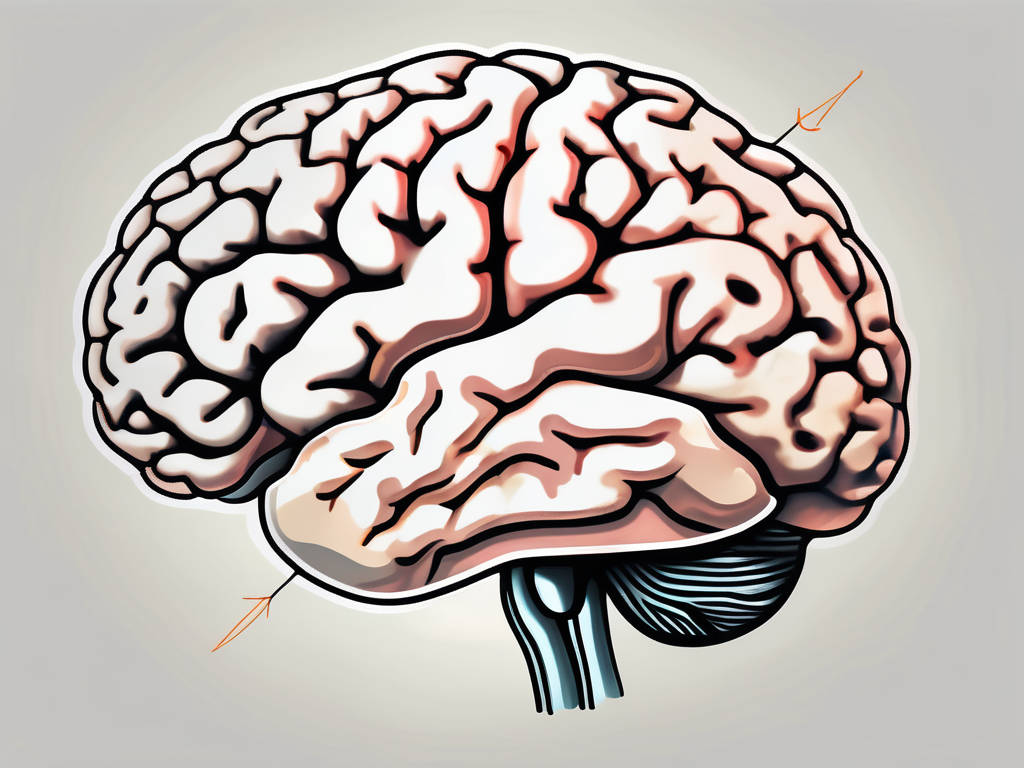
The trochlear nerve, also known as the fourth cranial nerve, plays a crucial role in our eye movements. It is responsible for controlling the superior oblique muscle, which helps in moving the eye in a specific direction. When this nerve is paralyzed or damaged, it can lead to various challenges and changes in our ability to control the movement of our eyes. In this article, we will explore the anatomy and function of the trochlear nerve, the impact of paralysis on this nerve, directional changes that may occur, treatment and management options, and how individuals can cope with living with trochlear nerve paralysis.
Understanding the Trochlear Nerve
The trochlear nerve is one of the twelve cranial nerves that emerge directly from the brain. It is unique because it is the only nerve that arises from the dorsal side of the brainstem, specifically the midbrain region. This nerve plays a crucial role in the movement and coordination of the eye.
Let’s delve deeper into the anatomy of the trochlear nerve to understand its structure and pathway.
Anatomy of the Trochlear Nerve
The trochlear nerve originates from the trochlear nucleus, which is located in the midbrain. It emerges from the brainstem near the cerebral peduncles and crosses over to the contralateral side before entering the cavernous sinus.
As it continues its journey, the trochlear nerve enters the orbit through the superior orbital fissure. Once inside the orbit, it innervates the superior oblique muscle, which is responsible for moving the eye downward and outward.
The superior oblique muscle plays a vital role in eye movement, allowing us to focus on objects positioned below our visual field. Its coordinated action with other eye muscles ensures proper alignment and binocular vision.
Function of the Trochlear Nerve
The trochlear nerve mainly controls the movement of the eye in a downward and outward direction. It aids in the rotation and depression of the eye, allowing us to shift our gaze and focus on objects at different angles.
Additionally, the superior oblique muscle helps in torsion, which is the rotation of the eye along its vertical axis. This rotational movement allows us to adjust our visual perception and maintain a clear line of sight.
When the trochlear nerve functions properly, it coordinates the movements of both eyes, ensuring proper alignment and binocular vision. This coordination is essential for depth perception and accurate visual interpretation of the world around us.
Understanding the trochlear nerve and its intricate role in eye movement provides valuable insights into the complexities of our visual system. The precise coordination of cranial nerves is crucial for our ability to navigate the world and perceive it in all its vivid detail.
The Impact of Paralysis on the Trochlear Nerve
Paralysis of the trochlear nerve can have significant effects on the individual’s ability to control eye movements and maintain visual alignment. It can result in impairments that affect daily activities and quality of life.
When the trochlear nerve, also known as the fourth cranial nerve, is paralyzed, it can lead to a condition called trochlear nerve palsy. This condition disrupts the normal functioning of the nerve, which is responsible for controlling the superior oblique muscle of the eye. The superior oblique muscle plays a crucial role in eye movements, particularly in looking downward and inward.
When the trochlear nerve is paralyzed, the affected individual may experience a range of symptoms. One of the most common symptoms is double vision, especially when looking downward. This occurs because the superior oblique muscle is unable to properly coordinate with the other muscles responsible for eye movements. As a result, the images from each eye do not align correctly, leading to double vision.
In addition to double vision, individuals with trochlear nerve paralysis may also experience upward deviation of the affected eye. This means that when they try to look straight ahead, the eye may drift upward, causing a misalignment with the unaffected eye. This misalignment can further contribute to the development of double vision and make it difficult for the individual to focus on objects.
Another common symptom of trochlear nerve paralysis is the inability to move the affected eye downward. This limitation in eye movement can make it challenging to perform tasks that require looking downward, such as reading or using electronic devices. Individuals may find themselves tilting their head or adjusting their body position to compensate for the impaired eye movements.
Causes of Trochlear Nerve Paralysis
Trochlear nerve paralysis can occur due to various factors, including trauma to the head or face, infections such as meningitis or encephalitis, brain tumors, vascular lesions, and congenital malformations. In some cases, the cause may be unknown.
Head or facial trauma can damage the trochlear nerve, leading to paralysis. This type of trauma can occur as a result of accidents, falls, or direct blows to the head or face. The force of the impact can disrupt the delicate structures of the nerve, causing it to lose its ability to transmit signals effectively.
Infections, such as meningitis or encephalitis, can also affect the trochlear nerve. These conditions involve inflammation of the meninges, which are the protective membranes surrounding the brain and spinal cord. The inflammation can extend to the nerves, including the trochlear nerve, and impair their normal functioning.
Brain tumors and vascular lesions can exert pressure on the trochlear nerve, leading to paralysis. Tumors can grow within the brain or near the nerve pathways, causing compression and damage. Vascular lesions, such as aneurysms or arteriovenous malformations, can disrupt the blood flow to the nerve, depriving it of essential nutrients and oxygen.
Congenital malformations, although rare, can also contribute to trochlear nerve paralysis. These malformations are present at birth and can affect the development and structure of the nerve. They may result from genetic factors or abnormal fetal development.
If you suspect you have trochlear nerve paralysis, it is important to consult with a healthcare professional who can conduct a thorough evaluation and provide an accurate diagnosis. They may perform a physical examination, review medical history, and order imaging tests to identify the underlying cause.
Symptoms and Diagnosis of Trochlear Nerve Paralysis
The symptoms of trochlear nerve paralysis can vary depending on the severity of the condition. Some common signs include double vision (especially when looking downward), upward deviation of the affected eye, inability to move the eye downward, difficulty reading, and tilting of the head to compensate for the impaired eye movements.
When individuals present with these symptoms, healthcare professionals can diagnose trochlear nerve paralysis through a comprehensive evaluation. They may assess eye movements, perform a thorough neurological examination, and utilize diagnostic imaging, such as magnetic resonance imaging (MRI), to visualize the affected area.
During the eye movement assessment, the healthcare professional will observe the individual’s ability to move their eyes in different directions, paying particular attention to any limitations or abnormalities. They may use specialized equipment to measure eye movements accurately and objectively.
The neurological examination involves assessing the function of the trochlear nerve and other cranial nerves. The healthcare professional will evaluate the individual’s ability to perform specific eye movements, such as looking upward, downward, and inward. They may also test the individual’s visual acuity and perform additional tests to assess the overall health of the eyes and surrounding structures.
In some cases, diagnostic imaging, such as MRI, may be necessary to visualize the trochlear nerve and identify any structural abnormalities or lesions. MRI uses powerful magnets and radio waves to create detailed images of the brain and surrounding tissues, allowing healthcare professionals to assess the condition of the nerve and determine the underlying cause of the paralysis.
Once a diagnosis of trochlear nerve paralysis is confirmed, healthcare professionals can develop an appropriate treatment plan tailored to the individual’s needs. Treatment options may include medication, vision therapy, or surgical interventions, depending on the underlying cause and severity of the paralysis.
Directional Changes Due to Trochlear Nerve Paralysis
Paralysis of the trochlear nerve can lead to significant directional changes in eye movements. Understanding these changes is crucial for individuals affected by trochlear nerve paralysis, as it can impact daily activities and overall visual perception.
Trochlear nerve paralysis, also known as fourth nerve palsy, is a condition that affects the ability to move the eye in a downward and outward direction. This nerve is responsible for controlling the superior oblique muscle, which is responsible for moving the eye downward and outward. When the trochlear nerve is paralyzed or damaged, the affected eye may have difficulty moving downward and may appear higher relative to the unaffected eye.
This can result in a condition known as hypertropia, where one eye appears higher than the other in primary gaze or when looking straight ahead. Individuals with trochlear nerve paralysis often experience vertical diplopia, where they see two images vertically offset from each other. This double vision can make it challenging to perform everyday tasks that require accurate depth perception, such as driving or playing sports.
Eye Movement and the Trochlear Nerve
The trochlear nerve plays a crucial role in coordinating eye movements. It is the smallest cranial nerve and has the longest intracranial course. It originates from the dorsal aspect of the midbrain and innervates the superior oblique muscle, which is responsible for moving the eye downward and outward.
When the trochlear nerve is functioning correctly, it allows for smooth and coordinated eye movements. However, when it is paralyzed or damaged, the affected eye’s ability to move downward is compromised, leading to a noticeable difference in eye alignment.
Individuals with trochlear nerve paralysis may experience difficulty looking down, making tasks such as reading or navigating stairs more challenging. They may need to tilt their head or adopt specific head positions to align their eyes properly and compensate for the impaired eye movements. However, these compensatory strategies may not be sufficient for all activities, and individuals may still experience difficulty with certain tasks that require precise eye movements.
How Paralysis Affects Directional Control
Trochlear nerve paralysis affects the individual’s ability to control the movement of their eyes accurately. The impaired downward and outward eye movements can disrupt the normal visual field and cause difficulties in daily activities.
For example, reading can become more challenging as the affected eye may struggle to move downward to track the lines of text. Individuals may need to use their finger or other aids to guide their reading and ensure they do not skip lines or lose their place.
Navigating stairs can also pose a problem for individuals with trochlear nerve paralysis. The downward gaze required to see the steps and judge their depth can be compromised, making it harder to navigate safely. This can lead to a higher risk of tripping or falling.
Furthermore, activities that involve precise eye movements, such as playing sports or engaging in hobbies that require hand-eye coordination, may become more challenging. The affected individual may struggle to track moving objects or accurately judge distances, impacting their performance and enjoyment of these activities.
It is important for individuals with trochlear nerve paralysis to work closely with healthcare professionals, such as ophthalmologists and occupational therapists, to develop strategies and adaptations that can help improve their functional abilities. These professionals can provide guidance on exercises, visual aids, and assistive devices that can enhance eye movements and mitigate the challenges associated with trochlear nerve paralysis.
Treatment and Management of Trochlear Nerve Paralysis
Trochlear nerve paralysis is a condition that requires careful management to address the underlying cause, manage symptoms, and maximize functional vision. Let’s explore the various treatment options available to individuals with trochlear nerve paralysis.
Medical Interventions for Trochlear Nerve Paralysis
The treatment approach for trochlear nerve paralysis depends on the underlying cause. Healthcare professionals may prescribe medications, such as anti-inflammatory drugs or antibiotics for infections, to alleviate symptoms and promote healing. In some cases, surgical interventions may be necessary to address structural abnormalities or remove tumors that are affecting the trochlear nerve.
When facing trochlear nerve paralysis, it is essential to consult with a healthcare professional who specializes in neurology or ophthalmology. They will conduct a thorough evaluation and diagnosis to determine the most appropriate treatment plan for your specific condition. By seeking expert guidance, you can receive personalized care and make informed decisions about your treatment options.
Rehabilitation and Therapy Options
Visual rehabilitation and therapy play a crucial role in managing trochlear nerve paralysis. Occupational therapists, optometrists, and ophthalmologists specializing in vision therapy can design customized programs to improve eye coordination, visual perception, and adaptation to the changes in eye movement.
These therapies may include exercises to strengthen the eye muscles, prism glasses to improve binocular vision, and techniques to promote visual perceptual skills. By actively participating in these therapies and following the guidance of healthcare professionals, individuals with trochlear nerve paralysis can enhance their visual capabilities and regain a sense of control over their daily activities.
Regular follow-ups with the healthcare professionals involved in your treatment are crucial. These appointments allow for progress tracking and adjustments to the treatment plan as necessary. By maintaining open communication with your healthcare team, you can ensure that your therapy remains effective and tailored to your evolving needs.
Remember, trochlear nerve paralysis management is a multidisciplinary effort that requires collaboration between healthcare professionals and the individual affected. With the right treatment and therapy, individuals with trochlear nerve paralysis can achieve improved visual function and enhance their overall quality of life.
Living with Trochlear Nerve Paralysis
Living with trochlear nerve paralysis can present unique challenges and require individuals to adapt to changes in their visual function. The trochlear nerve, also known as the fourth cranial nerve, plays a crucial role in controlling the movement of the superior oblique muscle of the eye. When this nerve is paralyzed, it can result in difficulty in moving the affected eye in a downward and inward direction.
Individuals with trochlear nerve paralysis may experience symptoms such as double vision, eye misalignment, and difficulty in reading or performing tasks that require downward eye movements. Coping with these directional changes can be challenging, but there are strategies that may help individuals adapt and improve their quality of life.
Coping Strategies for Directional Changes
Consulting with healthcare professionals who specialize in vision rehabilitation can provide valuable guidance and support in adapting to the directional changes caused by trochlear nerve paralysis. These professionals, such as optometrists or ophthalmologists, have expertise in assessing visual function and can assist in developing compensatory strategies.
One such strategy is the use of prisms. Prisms can be prescribed to individuals with trochlear nerve paralysis to help shift the image seen by the affected eye, thereby improving alignment and reducing double vision. These prisms can be incorporated into glasses or contact lenses, providing a practical and convenient solution for managing visual symptoms.
Additionally, making adjustments to the environment can be helpful. Ensuring proper lighting can reduce eye strain and make it easier to focus on objects. Using larger fonts for reading materials can alleviate the strain on the eyes and make reading more comfortable. Organizing items in a way that minimizes the need for frequent downward eye movements can also make daily activities more manageable.
Long-Term Prognosis and Quality of Life
The long-term prognosis for trochlear nerve paralysis varies depending on the underlying cause and individual factors. In some cases, trochlear nerve paralysis may resolve with appropriate treatment, such as addressing the underlying condition causing the paralysis. However, in other cases, ongoing management strategies may be necessary to optimize functional vision.
Regular follow-ups with healthcare professionals are essential for individuals with trochlear nerve paralysis. These follow-ups allow for monitoring of visual function and adjustment of treatment plans as necessary. It is important to communicate any ongoing symptoms or changes in visual function to healthcare professionals, as they can provide ongoing support and make necessary adjustments to promote the highest possible quality of life.
In conclusion, trochlear nerve paralysis can significantly impact the directional control of eye movements. Understanding the anatomy and function of the trochlear nerve, the causes and symptoms of paralysis, and the available treatment and management options is crucial for individuals affected by this condition. By working closely with healthcare professionals and utilizing rehabilitation and therapy strategies, individuals can navigate the challenges posed by trochlear nerve paralysis and optimize their functional vision. With the right support and strategies in place, individuals can lead fulfilling lives despite the challenges presented by trochlear nerve paralysis.
