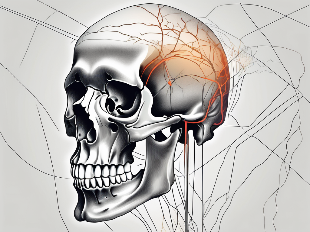
The trochlear nerve is a crucial component of our nervous system, responsible for providing motor innervation to the superior oblique muscle of the eye. To fully understand the significance of this nerve, it is essential to explore its anatomy, function, and the specific foramen it traverses. Additionally, we will delve into the implications of trochlear nerve damage, address common misconceptions, and provide further resources for a comprehensive understanding of this intricate topic.
Understanding the Trochlear Nerve
Anatomy of the Trochlear Nerve
The trochlear nerve, also known as the fourth cranial nerve, plays a vital role in controlling eye movements. It arises from the trochlear nucleus, located in the midbrain, and travels in a distinctive course. Emerging from the dorsal surface of the brainstem, the trochlear nerve courses along the lateral wall of the cavernous sinus, which is a dural venous sinus located on either side of the sella turcica. This distinctive pathway sets it apart from the other cranial nerves.
The trochlear nerve then courses through the superior orbital fissure, a narrow passage located in the sphenoid bone. By passing through this foramen, the nerve gains access to the orbit and innervates the superior oblique muscle, which is responsible for various eye movements.
As the trochlear nerve traverses through the superior orbital fissure, it is surrounded by various structures that contribute to the complex anatomy of the orbit. These structures include the ophthalmic artery, which supplies blood to the eye, and the oculomotor nerve, which controls the movements of several other eye muscles.
Furthermore, the trochlear nerve’s path through the cavernous sinus exposes it to potential anatomical variations and clinical implications. The cavernous sinus houses important structures such as the internal carotid artery and the abducens nerve, making it a critical region for understanding the trochlear nerve’s function and potential pathologies.
Function of the Trochlear Nerve
The trochlear nerve primarily controls the superior oblique muscle, one of the six extraocular muscles responsible for eye movement. The synergistic action of these muscles enables us to gaze in different directions, track moving objects, and maintain equilibrium.
Specifically, the trochlear nerve’s innervation of the superior oblique muscle facilitates downward, outward, and inward eye movements. It plays a crucial role in allowing us to look down while tilting our head, a movement essential for activities such as reading or walking downstairs.
In addition to its role in eye movements, the trochlear nerve also contributes to the coordination and alignment of both eyes. By working in conjunction with the other cranial nerves responsible for eye movements, the trochlear nerve ensures that both eyes move in a synchronized manner, allowing for binocular vision and depth perception.
Disorders affecting the trochlear nerve can lead to a condition known as trochlear nerve palsy, which results in a variety of symptoms. These symptoms may include double vision, difficulty looking downward or inward, and a head tilt to compensate for the affected eye’s limited movement. Understanding the anatomy and function of the trochlear nerve is crucial for diagnosing and managing such conditions.
The Role of Foramina in the Human Body
Definition and Function of Foramina
Foramina, plural foramen, are essential structures in the human body that allow the passage of various anatomical components. These minute openings are found in bones, serving as conduits for blood vessels, nerves, and other vital structures.
Foramina play a vital role in maintaining the intricate network of connections throughout our body. They provide pathways for nerves to innervate specific regions, ensuring optimal functioning of bodily systems.
Foramina are not only crucial for the passage of nerves and blood vessels but also play a significant role in the distribution of nutrients and oxygen. These tiny openings allow blood vessels to deliver oxygen-rich blood to different parts of the body, ensuring proper nourishment and functioning of cells.
In addition to their role in the circulatory system, foramina also facilitate the removal of waste products and toxins from tissues. Through these openings, waste materials are transported away from cells, promoting overall health and well-being.
Furthermore, foramina contribute to the body’s ability to regulate temperature. By allowing blood vessels to dilate or constrict, these openings help maintain optimal body temperature, ensuring that organs and tissues function efficiently.
Major Foramina and Their Roles
While the human body harbors numerous foramina, several hold particular importance. The superior orbital fissure is one such critical foramen.
Multiple cranial nerves pass through the superior orbital fissure, including the trochlear nerve. This foramen serves as the gateway for these nerves to enter the orbit, enabling essential motor and sensory functions within the eye.
In addition to the superior orbital fissure, the foramen magnum is another significant opening in the human body. Located at the base of the skull, this large foramen allows the spinal cord to pass through and connect with the brain. It serves as a vital pathway for the transmission of sensory and motor information between the brain and the rest of the body.
Another notable foramen is the obturator foramen, located in the pelvic region. This large opening allows for the passage of nerves, blood vessels, and muscles that are essential for the movement and stability of the lower limbs. Without the obturator foramen, normal walking and other lower limb activities would be compromised.
Moreover, the jugular foramen, situated at the base of the skull, plays a crucial role in the drainage of blood from the brain. This foramen allows the internal jugular vein to exit the skull and return deoxygenated blood back to the heart. Without the jugular foramen, the brain would be unable to efficiently remove waste products and maintain proper blood flow.
These are just a few examples of the many foramina found in the human body, each with its unique role in ensuring the proper functioning of various bodily systems. From facilitating nerve transmission to enabling blood circulation and nutrient distribution, foramina are indispensable structures that contribute to our overall health and well-being.
The Specific Foramen for the Trochlear Nerve
Identifying the Trochlear Nerve’s Foramen
To comprehend the specific foramen through which the trochlear nerve passes, we must recognize the importance of the superior orbital fissure. This foramen, situated within the sphenoid bone, channels the trochlear nerve into the orbit.
The superior orbital fissure is a narrow, elongated opening located at the junction of the greater and lesser wings of the sphenoid bone. It serves as a crucial pathway for several structures, including the trochlear nerve, as they make their way into the orbit. The superior orbital fissure is bordered by the frontal, sphenoid, and ethmoid bones, forming a protective bony canal.
Identification of the superior orbital fissure is crucial in understanding the trochlear nerve’s route and its potential vulnerabilities. By studying the anatomical landmarks and structures surrounding this foramen, medical professionals can gain valuable insights into the potential impact of trauma or pathology on the trochlear nerve’s function.
The Pathway of the Trochlear Nerve through the Foramen
As the trochlear nerve courses through the superior orbital fissure, it enters the orbit and carries vital motor innervation for the superior oblique muscle. This pathway allows precise control over eye movements, contributing to our ability to focus and track objects effectively.
The superior oblique muscle plays a crucial role in eye movement by rotating the eyeball downward and outward. This movement is essential for maintaining proper alignment and coordination of both eyes, allowing us to accurately perceive depth and distance. The trochlear nerve, also known as the fourth cranial nerve, supplies the superior oblique muscle with the necessary signals to carry out these precise movements.
Given the significance of this foramen and its role in facilitating the trochlear nerve’s passage, any potential damage or impingement can have significant consequences on eye movement and coordination. Trauma, tumors, or inflammation affecting the superior orbital fissure can lead to trochlear nerve dysfunction, resulting in diplopia (double vision), difficulty in looking downward, and an overall decrease in eye movement control.
In conclusion, understanding the specific foramen for the trochlear nerve, namely the superior orbital fissure, is crucial for comprehending the nerve’s pathway and its role in eye movement. By recognizing the anatomical details and potential vulnerabilities associated with this foramen, healthcare professionals can better diagnose and treat conditions affecting the trochlear nerve, ultimately improving patients’ visual function and quality of life.
Implications of Trochlear Nerve Damage
The trochlear nerve, also known as the fourth cranial nerve, plays a crucial role in eye movement. Damage to this nerve can have significant implications on a person’s vision and overall quality of life. Understanding the symptoms and treatment options for trochlear nerve damage is essential for prompt diagnosis and effective management.
Symptoms of Trochlear Nerve Damage
Trochlear nerve damage can manifest in various ways, leading to distinct symptoms. One of the most common symptoms is diplopia, also known as double vision. This occurs when the eyes are unable to align properly, resulting in seeing two images instead of one. Vertical misalignment of the eyes is another symptom, where one eye appears higher or lower than the other. This can cause difficulty in focusing or tracking objects, making everyday tasks challenging. Additionally, individuals with trochlear nerve damage may experience frequent headaches or eye strain due to the extra effort required to see clearly.
If you experience any of these symptoms, it is crucial to consult with a qualified healthcare professional for a comprehensive evaluation. Proper diagnosis is essential to determine the underlying cause of trochlear nerve damage and develop an appropriate treatment plan.
Treatment and Recovery for Trochlear Nerve Damage
When it comes to trochlear nerve damage, prompt diagnosis and personalized treatment are essential for a successful recovery. The specific approach will depend on the underlying cause and severity of the impairment.
Treatment options for trochlear nerve damage may include conservative management techniques. These can involve the use of eye patching or prism glasses to correct vision distortions and alleviate double vision. Physical therapy is another valuable treatment option, focusing on enhancing eye movements and coordination. Through targeted exercises and techniques, individuals can improve their ability to control and align their eyes properly.
In severe cases or when there are structural abnormalities affecting the trochlear nerve, surgical interventions may be necessary. These procedures aim to repair or restore the damaged nerve, allowing for improved eye movement and function.
Regardless of the chosen treatment approach, it is vital to follow medical advice and attend regular follow-up appointments to monitor progress and ensure optimal recovery. The recovery process for trochlear nerve damage can vary from person to person, and close monitoring is necessary to make any necessary adjustments to the treatment plan.
In conclusion, trochlear nerve damage can have significant implications on a person’s vision and daily life. Understanding the symptoms and seeking prompt medical attention is crucial for accurate diagnosis and effective treatment. With the right approach and proper care, individuals with trochlear nerve damage can experience improved vision and a better quality of life.
Frequently Asked Questions about the Trochlear Nerve and Foramen
Common Misconceptions about the Trochlear Nerve
One common misconception is assuming that trochlear nerve damage will always cause pain or discomfort. While some individuals may experience headaches or eye strain, others may not report any physical sensations. Therefore, it is crucial to recognize that symptoms can vary among individuals.
It is important to note that the trochlear nerve, also known as the fourth cranial nerve, is responsible for the movement of the superior oblique muscle in the eye. This muscle helps to control eye movements, particularly when looking down and inward. Damage to the trochlear nerve can result in a condition called trochlear nerve palsy, which can lead to double vision and difficulty with downward eye movements.
In addition to pain and discomfort, trochlear nerve damage can also cause other symptoms such as blurred vision, difficulty focusing, and eye misalignment. These symptoms can significantly impact a person’s daily life and may require medical intervention.
It is worth mentioning that trochlear nerve damage can occur due to various reasons, including trauma, infections, tumors, and vascular disorders. Understanding the underlying cause of the nerve damage is essential for proper diagnosis and treatment.
Further Resources for Understanding the Trochlear Nerve and Foramen
For individuals seeking additional information and a deeper understanding of the trochlear nerve and related topics, consulting reputable medical resources is highly recommended. Qualified healthcare professionals, medical textbooks, and reliable online platforms dedicated to anatomy and neurology can provide valuable insights.
Exploring medical textbooks that focus on neuroanatomy can provide a comprehensive understanding of the trochlear nerve’s structure, function, and clinical significance. These resources often include detailed illustrations and explanations, making it easier to grasp complex concepts.
Furthermore, seeking guidance from healthcare professionals specializing in neurology or ophthalmology can offer personalized information and address specific concerns related to the trochlear nerve. These professionals can provide accurate diagnoses, treatment options, and recommendations for managing trochlear nerve-related conditions.
Remember, this article serves as a general overview, and professional medical advice should always be sought when addressing specific concerns or conditions related to the trochlear nerve and associated foramen.
