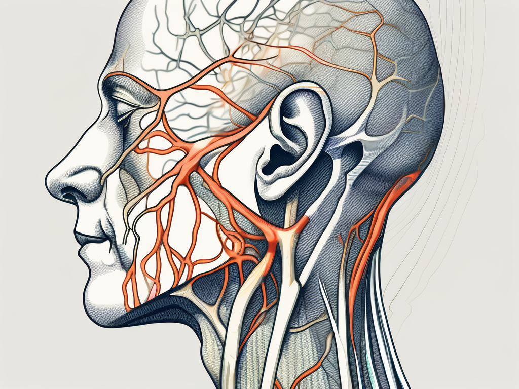
The trochlear nerve, also known as the fourth cranial nerve, plays a crucial role in the movement of the eye. When this nerve is damaged or impaired, it can lead to a variety of symptoms, including head tilt. In this article, we will explore the anatomy and function of the trochlear nerve, the impact of trochlear nerve lesion, the mechanism and variations of head tilt in trochlear nerve lesion, diagnosis techniques, treatment options, and prognosis for patients with this condition.
Understanding the Trochlear Nerve
The trochlear nerve, also known as cranial nerve IV, is one of the twelve pairs of cranial nerves in the human body. It plays a crucial role in coordinating eye movements and is responsible for the innervation of the superior oblique muscle. Let’s delve deeper into the fascinating world of the trochlear nerve and explore its anatomy and function.
Anatomy of the Trochlear Nerve
The trochlear nerve originates in the midbrain, specifically the posterior aspect known as the dorsal midbrain. It emerges from the brainstem and takes a unique and intriguing path. Unlike other cranial nerves, it travels dorsally, meaning it moves towards the back of the brain, before crossing over to the opposite side. This distinctive course makes the trochlear nerve susceptible to certain forms of injury.
As the trochlear nerve continues its journey, it courses along the lateral wall of a structure called the cavernous sinus. The cavernous sinus is a complex network of veins and nerves located on either side of the sella turcica, a bony saddle-shaped structure that houses the pituitary gland. This intricate pathway adds to the trochlear nerve’s complexity and highlights its importance.
After traversing the cavernous sinus, the trochlear nerve enters the orbit, or eye socket, through a small opening called the superior orbital fissure. Once inside the orbit, it innervates the superior oblique muscle, which is responsible for specific eye movements.
Function of the Trochlear Nerve
The trochlear nerve’s primary function is to control the superior oblique muscle, a small but mighty muscle that plays a significant role in eye movements. The superior oblique muscle is responsible for various actions, including depression (downward movement), intorsion (inward rotation), and abduction (outward movement) of the eye.
When the trochlear nerve is functioning properly, these movements are smooth, precise, and coordinated. This coordination allows the eyes to track objects accurately, contributing to our visual perception and depth perception. The trochlear nerve’s intricate control over the superior oblique muscle is essential for maintaining optimal eye function.
It is worth noting that the trochlear nerve’s long intracranial course and unique pathway make it vulnerable to certain forms of injury. Trauma, tumors, or other pathological conditions affecting the midbrain or cavernous sinus can potentially impact the trochlear nerve’s function and lead to eye movement abnormalities.
In conclusion, the trochlear nerve is a remarkable cranial nerve that plays a vital role in coordinating eye movements. Its anatomy, with its dorsal path and passage through the cavernous sinus, adds to its intrigue. Understanding the trochlear nerve’s function and its potential vulnerabilities can help us appreciate the complexity of our visual system and the delicate balance required for optimal eye function.
The Impact of Trochlear Nerve Lesion
A trochlear nerve lesion can lead to a range of symptoms and complications, with head tilt being one of the most notable signs. However, it is important to recognize that head tilt alone is not exclusive to trochlear nerve lesions and can present in other conditions as well. Consultation with a healthcare professional is essential to determine the underlying cause of head tilt and provide appropriate treatment.
When a trochlear nerve lesion occurs, it can have a significant impact on a person’s vision and overall quality of life. The trochlear nerve, also known as the fourth cranial nerve, is responsible for controlling the movement of the superior oblique muscle in the eye. This muscle plays a crucial role in eye movements, particularly when looking downward or inward.
Symptoms Associated with Trochlear Nerve Lesion
In addition to head tilt, patients with trochlear nerve lesions may experience double vision (diplopia), particularly when looking downward or inward. This occurs due to the impaired function of the superior oblique muscle, leading to an imbalance in eye movements. The brain receives conflicting signals from the affected eye, resulting in the perception of two images instead of one.
Double vision can be extremely disorienting and can significantly impact a person’s ability to perform daily activities such as reading, driving, or even walking. Patients may find it challenging to focus on objects or may experience difficulty judging distances accurately. This can lead to eye strain, headaches, and overall visual discomfort.
Furthermore, trochlear nerve lesions can also affect visual acuity, causing a decrease in the clarity and sharpness of vision. Patients may notice that their vision becomes blurry or that they have difficulty reading small print. This can further contribute to eye strain and may require the use of corrective lenses or other visual aids.
Causes and Risk Factors of Trochlear Nerve Lesion
Trochlear nerve lesions can be caused by various factors, including trauma, tumors, infections, and vascular disorders. Direct head trauma or skull fractures can damage the nerve directly or indirectly by compressing the surrounding structures. For example, a severe blow to the head during a car accident or a fall can result in nerve damage, leading to trochlear nerve lesion.
Tumors, such as meningiomas or gliomas, can also exert pressure on the nerve, leading to its dysfunction. These tumors can develop in the brain or in the structures surrounding the nerve, causing compression and interference with its normal function. Early detection and treatment of these tumors are crucial to prevent further damage to the trochlear nerve and preserve vision.
Infections, such as meningitis or encephalitis, can also affect the trochlear nerve. These infections can cause inflammation in the brain and surrounding structures, leading to nerve damage. Prompt treatment with antibiotics or antiviral medications is essential to minimize the risk of permanent nerve damage and associated complications.
Additionally, certain systemic conditions, such as diabetes or hypertension, may increase the risk of trochlear nerve lesions. These conditions can affect the blood vessels supplying the nerve, leading to reduced blood flow and oxygenation. Over time, this can result in nerve damage and dysfunction.
In conclusion, trochlear nerve lesions can have a significant impact on a person’s vision and overall well-being. It is important to recognize the symptoms associated with trochlear nerve lesions, such as head tilt and double vision, and seek medical attention for proper diagnosis and treatment. Early intervention can help minimize the complications associated with trochlear nerve lesions and improve the patient’s quality of life.
Head Tilt in Trochlear Nerve Lesion
The mechanism of head tilt in trochlear nerve lesion is quite intriguing. The superior oblique muscle, controlled by the trochlear nerve, normally functions to depress and intort the eye. However, when the nerve is impaired, the opposing muscles, such as the inferior oblique muscle, are no longer counteracted, resulting in an unopposed contraction and head tilt. This compensatory mechanism aims to align the visual axes, reducing double vision and improving visual comfort.
Mechanism of Head Tilt in Trochlear Nerve Lesion
When the trochlear nerve is damaged on one side, the superior oblique muscle on the affected side weakens. To compensate for this weakness, the patient tilts their head in the opposite direction of the affected eye, allowing the visual axes to align more closely. By tilting the head, the patient minimizes the vertical misalignment of the eyes, reducing diplopia and improving visual clarity.
Moreover, the head tilt also helps to alleviate the strain on the weakened superior oblique muscle. By tilting the head, the patient reduces the amount of work required by the impaired muscle to align the visual axes. This reduction in muscle effort can provide relief and prevent further strain on the weakened muscle, promoting healing and recovery.
Furthermore, the head tilt in trochlear nerve lesion can have additional effects on the patient’s posture and body alignment. Tilting the head to one side may cause a compensatory shift in the rest of the body, as the patient tries to maintain balance and stability. This shift in body alignment can involve subtle adjustments in the spine, hips, and shoulders, as well as changes in weight distribution.
Variations in Head Tilt Direction
Although head tilt is a common feature of trochlear nerve lesions, the direction of the tilt can vary among individuals. Some patients may tilt their head towards the affected side, while others may tilt it away from the affected side. The direction of the head tilt is influenced by various factors, such as the severity and chronicity of the lesion, as well as individual compensatory mechanisms.
It is important to note that the direction of the head tilt does not necessarily indicate the severity or prognosis of the trochlear nerve lesion. The variation in tilt direction is primarily a result of individual adaptation and compensation strategies. Therefore, the direction of the head tilt should be carefully assessed and considered in conjunction with other clinical findings to determine the underlying cause and appropriate management of the trochlear nerve lesion.
Additionally, the direction of the head tilt may also be influenced by factors such as visual dominance and handedness. Some studies suggest that individuals with right-handedness and right-eye dominance may be more likely to tilt their head towards the affected side, while those with left-handedness and left-eye dominance may tilt their head away from the affected side. These observations highlight the complex interplay between neurological, visual, and motor factors in determining the direction of head tilt in trochlear nerve lesions.
Diagnosis of Trochlear Nerve Lesion
Accurate diagnosis of trochlear nerve lesion is crucial for appropriate management and treatment. Healthcare professionals utilize a combination of clinical examination techniques and imaging studies to evaluate patients suspected of having a trochlear nerve lesion.
Trochlear nerve lesions can present with a variety of symptoms, including diplopia (double vision), vertical misalignment of the eyes, and difficulty in looking downwards. To determine the presence and severity of a trochlear nerve lesion, healthcare professionals employ a range of clinical examination techniques.
Clinical Examination Techniques
During the clinical examination, the healthcare professional will assess eye movements, including the alignment and range of motion. Special attention is given to the superior oblique muscle function, which is crucial for the diagnosis of trochlear nerve lesions.
The Parks-Bielschowsky three-step test is a commonly used clinical examination technique. This test involves the patient tilting their head in different directions while focusing on a target. By observing the pattern of eye movement and the presence of any vertical misalignment, healthcare professionals can gain insight into the functioning of the trochlear nerve.
Another clinical examination technique used is the head tilt test. In this test, the patient is asked to tilt their head to one side while focusing on a target. The healthcare professional carefully observes any compensatory head tilt or abnormal eye movements, which can indicate a trochlear nerve lesion.
Imaging and Other Diagnostic Tools
Imaging studies, such as magnetic resonance imaging (MRI) or computed tomography (CT) scans, are essential for visualizing the brainstem, cranial nerves, and associated structures. These imaging modalities help identify any structural abnormalities or lesions that may be affecting the trochlear nerve.
MRI scans provide detailed images of the brain and cranial nerves, allowing healthcare professionals to visualize the trochlear nerve and surrounding structures. CT scans, on the other hand, provide cross-sectional images that can help identify bony abnormalities or fractures that may be compressing the trochlear nerve.
In certain cases, electrophysiological testing, such as electromyography, may be used to measure the electrical activity and response of the affected muscle. This can help determine the extent of nerve damage and provide additional information for diagnosis and treatment planning.
Overall, the diagnosis of trochlear nerve lesion requires a comprehensive approach that combines clinical examination techniques and imaging studies. By carefully evaluating the patient’s symptoms, conducting thorough examinations, and utilizing advanced diagnostic tools, healthcare professionals can accurately diagnose trochlear nerve lesions and develop appropriate management and treatment strategies.
Treatment and Management of Trochlear Nerve Lesion
The treatment and management of trochlear nerve lesions depend on the underlying cause, severity of symptoms, and individual patient factors. It is important to consult with a healthcare professional to determine the appropriate course of action.
Trochlear nerve lesions can be challenging to treat due to the complex nature of the nerve and its role in eye movement. However, advancements in medical science have provided various treatment options to help alleviate symptoms and improve the quality of life for affected individuals.
Non-Surgical Treatment Options
In some cases, conservative management approaches may be sufficient to alleviate symptoms. This can include the use of prism glasses or other optical aids to correct double vision and enhance visual alignment. Prism glasses work by bending light, allowing the eyes to work together more effectively and reducing the strain on the trochlear nerve.
Physical therapy or exercises aimed at strengthening the surrounding muscles and improving eye movement coordination may also be beneficial. These exercises can help train the eyes to work together more efficiently, reducing the strain on the trochlear nerve and improving overall eye function.
Additionally, the use of botulinum toxin injections in certain situations can help relax overactive muscles and achieve better eye alignment. Botulinum toxin, commonly known as Botox, works by temporarily paralyzing specific muscles, allowing for better control and alignment of the eyes.
Surgical Interventions for Trochlear Nerve Lesion
Surgery is considered in cases where conservative management approaches fail to provide adequate relief or in the presence of specific structural abnormalities. Surgical interventions may involve repairing or replacing damaged portions of the trochlear nerve, decompressing the nerve from surrounding impinging structures, or addressing any associated tumors or vascular abnormalities.
The decision to undergo surgery is not taken lightly, as it carries its own risks and considerations. However, in certain cases, surgical intervention can provide significant improvements in symptoms and overall eye function.
The type of surgery performed will depend on the underlying cause and individual patient needs. Some common surgical procedures for trochlear nerve lesions include trochleoplasty, which involves reshaping the bony groove that the trochlear nerve passes through, and neurectomy, which involves the removal of the damaged portion of the nerve.
It is important to note that the success of surgical interventions for trochlear nerve lesions can vary depending on the individual case. Factors such as the extent of nerve damage, the presence of other underlying conditions, and the overall health of the patient can influence the outcome of the surgery.
Recovery from surgical interventions for trochlear nerve lesions can also vary. Some individuals may experience immediate improvements in symptoms, while others may require a longer period of rehabilitation and therapy to regain optimal eye function.
In conclusion, the treatment and management of trochlear nerve lesions require a comprehensive approach that takes into account the individual patient’s needs and the underlying cause of the condition. With the help of healthcare professionals and advancements in medical science, individuals with trochlear nerve lesions can find relief and improve their quality of life.
Prognosis and Recovery from Trochlear Nerve Lesion
Prognosis for patients with trochlear nerve lesion can vary depending on several factors, such as the extent and duration of the lesion, the underlying cause, and the overall health of the patient. While some individuals may experience partial or complete recovery of trochlear nerve function, others may have long-term deficits. Regular follow-up visits with a healthcare professional are essential to monitor progress, adjust treatment plans if necessary, and optimize visual outcomes.
Factors Influencing Recovery
Factors that may influence recovery from trochlear nerve lesions include the age of the patient, the duration of symptoms before treatment initiation, the underlying cause, and the effectiveness of the chosen treatment approach. Additionally, the presence of any associated complications, such as double vision, may affect the overall visual prognosis.
Long-Term Outlook for Patients with Trochlear Nerve Lesion
While living with a trochlear nerve lesion can present challenges, many patients are able to manage their symptoms effectively with appropriate treatment and support. It is essential to work closely with a healthcare professional to develop a tailored treatment plan and to address any concerns or difficulties that may arise. By taking proactive steps, individuals with trochlear nerve lesions can lead fulfilling lives and maintain good visual function.
