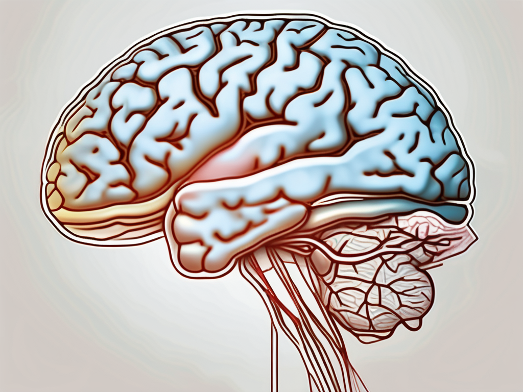
The trochlear nerve, also known as cranial nerve IV, plays a crucial role in visual movement and coordination. Understanding the anatomy and function of this nerve is essential for diagnosing and treating related disorders. In this article, we will explore the pathway of the trochlear nerve through the brainstem, along with its clinical importance and available treatment options.
Understanding the Trochlear Nerve
The trochlear nerve is one of the twelve cranial nerves originating from the brainstem. It is the smallest of these nerves and has a unique anatomical pathway. To comprehend its importance, let us delve into a detailed understanding of its anatomy and function.
Anatomy of the Trochlear Nerve
The trochlear nerve emerges from the dorsal aspect of the midbrain, near the inferior colliculus. It then curves around the brainstem and enters the cavernous sinus, a crucial structure responsible for transporting multiple cranial nerves. Continuing its journey, the trochlear nerve emerges from the cavernous sinus and enters the orbit through the superior orbital fissure.
Within the orbit, the trochlear nerve innervates the superior oblique muscle of the eye, which plays a crucial role in controlling vertical eye movements. Its positioning on the contralateral aspect of the midbrain is unique among the cranial nerves.
The trochlear nerve’s pathway through the brainstem is an intricate and fascinating journey. As it emerges from the midbrain, it navigates through various structures, each with its own significance. The dorsal aspect of the midbrain, where the trochlear nerve originates, is a region involved in the processing of auditory and visual information. This connection highlights the close relationship between the trochlear nerve and visual function.
Curving around the brainstem, the trochlear nerve encounters other cranial nerves, forming a network of communication and coordination. The cavernous sinus, through which the trochlear nerve passes, is a complex and vital structure. It houses not only the trochlear nerve but also the oculomotor, abducens, and trigeminal nerves, among others. This proximity allows for efficient transmission of signals and coordination of eye movements.
Upon exiting the cavernous sinus, the trochlear nerve enters the orbit through the superior orbital fissure. The orbit, also known as the eye socket, is a bony cavity that protects and houses the eyeball. Within this confined space, the trochlear nerve takes on its role in controlling the superior oblique muscle, contributing to the intricate dance of eye movements.
Function of the Trochlear Nerve
The primary function of the trochlear nerve is to control the superior oblique muscle, allowing for downward and inward eye movements. By coordinating the intricate movements of the eye, the trochlear nerve aids in proper visual alignment and depth perception.
Imagine looking up at the stars on a clear night. As you gaze upward, your eyes move in a smooth and coordinated manner, thanks to the trochlear nerve. It ensures that your eyes can effortlessly track celestial objects, allowing you to appreciate the beauty of the night sky.
Damage to the trochlear nerve can result in a condition called trochlear nerve palsy, characterized by difficulty with eye movements, specifically downward and inward rotations. This condition can significantly impact daily activities, such as reading, driving, and even walking down stairs. It highlights the crucial role the trochlear nerve plays in our visual function.
As we explore the clinical importance of the trochlear nerve’s pathway through the brainstem, it is crucial to understand the potential impact of such damage. By gaining a deeper understanding of the trochlear nerve’s anatomy and function, we can appreciate the intricate mechanisms that allow us to perceive the world around us and navigate through it with ease.
The Brainstem and Its Significance
The brainstem is a vital component of the central nervous system, connecting the brain to the spinal cord. It houses several vital structures responsible for regulating various bodily functions, including heart rate, breathing, and consciousness.
The brainstem is a complex and intricate region of the brain that plays a crucial role in maintaining homeostasis and coordinating various physiological processes. It is often referred to as the “bridge” between the brain and the rest of the body.
Anatomy of the Brainstem
The brainstem consists of three main regions: the midbrain, pons, and medulla oblongata. Each region plays a distinct role in relaying sensory and motor information to and from the brain and spinal cord.
The midbrain, located at the top of the brainstem, is responsible for controlling visual and auditory reflexes, as well as regulating eye movement. It also plays a crucial role in the production of dopamine, a neurotransmitter involved in reward and motivation.
The pons, located below the midbrain, serves as a bridge connecting various parts of the brain. It plays a vital role in regulating sleep, arousal, and facial movements. Additionally, it contains important nuclei involved in the control of breathing.
The medulla oblongata, located at the base of the brainstem, is responsible for controlling vital functions such as heart rate, blood pressure, and respiration. It also houses important nuclei involved in swallowing, coughing, and vomiting.
Within the brainstem, there are numerous pathways and connections that allow for the transmission of information between different parts of the brain and spinal cord. One particularly intriguing pathway is that of the trochlear nerve.
The trochlear nerve’s pathway through the brainstem is particularly intriguing due to its decussation, or crossing, at the level of the midbrain. This unique arrangement allows the trochlear nerve to control the contralateral superior oblique muscle. This muscle is responsible for downward and outward eye movement.
Role of the Brainstem in the Nervous System
The brainstem serves as a vital relay station for sensory and motor pathways in the nervous system. It plays a crucial role in regulating involuntary functions and coordinating voluntary movements.
One of the most fascinating aspects of the brainstem is its ability to regulate basic bodily functions without conscious effort. For example, it controls the rhythm and depth of breathing, ensuring that oxygen is delivered to the body’s tissues and carbon dioxide is removed.
Furthermore, the brainstem is involved in maintaining consciousness and alertness. It receives sensory information from various parts of the body and relays it to the cerebral cortex, allowing us to perceive and respond to our environment.
Understanding the intricate relationship between the trochlear nerve and the brainstem is pivotal in comprehending the impact of damage along its pathway. Let us now explore the precise points at which this nerve originates, exits the brainstem, and traverses its unique course.
The brainstem is a marvel of nature, with its intricate structure and vital functions. It is a testament to the complexity and beauty of the human body, and its significance cannot be overstated. Without the brainstem, our bodies would not be able to perform essential functions necessary for survival.
In conclusion, the brainstem is a remarkable part of the central nervous system that plays a crucial role in regulating various bodily functions. Its anatomy and function are intricately linked, allowing for the coordination of sensory and motor information. The trochlear nerve’s pathway through the brainstem is just one example of the fascinating connections within this region. Further research and exploration of the brainstem will undoubtedly continue to reveal its importance in maintaining homeostasis and overall well-being.
Path of the Trochlear Nerve Through the Brainstem
The trochlear nerve’s pathway through the brainstem involves intricate anatomical landmarks and crossing points. Exploring these details will provide a comprehensive understanding of the nerve’s journey and its potential clinical implications.
The trochlear nerve, also known as the fourth cranial nerve, has a unique and fascinating route through the brainstem. Let’s delve deeper into the origin, exit points, and pathway of this important nerve.
Origin and Exit Points of the Trochlear Nerve
As mentioned earlier, the trochlear nerve arises from the dorsal aspect of the midbrain, near the inferior colliculus. This positioning places it within close proximity to various other cranial nerves, emphasizing the complexity of its pathway.
Emerging from the midbrain, the trochlear nerve takes a distinct path. It travels caudally, curving around the contralateral cerebral peduncle. This unique positioning allows for its decussation, or crossing, which is a defining characteristic of the trochlear nerve’s pathway.
The trochlear nerve’s decussation is an intriguing feature. It means that the nerve fibers originating from one side of the brainstem cross over to the opposite side, resulting in a contralateral innervation pattern. This anatomical arrangement has significant implications for the control of eye movements and coordination.
Trochlear Nerve’s Pathway and Crossing Point
After the trochlear nerve exits the brainstem, it continues its journey through various structures in the head and neck. One important landmark it encounters is the cavernous sinus, a complex network of veins located on either side of the sella turcica, a bony structure in the skull.
Passing through the cavernous sinus, the trochlear nerve eventually reaches the superior orbital fissure. This narrow opening in the skull serves as a conduit for several cranial nerves, including the trochlear nerve, as they make their way into the orbit.
Upon reaching the orbit, the trochlear nerve innervates the superior oblique muscle, one of the extraocular muscles responsible for controlling eye movements. Its decussation enables it to influence the contralateral eye, further highlighting the intricacies of the nervous system.
The trochlear nerve’s pathway and crossing point play a crucial role in the coordination of eye movements. Dysfunction or damage to this nerve can result in a condition known as trochlear nerve palsy, characterized by weakness or paralysis of the superior oblique muscle. This can lead to various visual disturbances and difficulties in performing certain eye movements.
Understanding the detailed anatomy and pathway of the trochlear nerve is essential for clinicians and researchers in diagnosing and managing conditions related to this nerve. The complex interplay between the brainstem, cranial nerves, and ocular muscles highlights the remarkable precision and organization of the human nervous system.
Clinical Importance of the Trochlear Nerve’s Path
Understanding the course and functions of the trochlear nerve is crucial when assessing and managing related clinical conditions. Disorders affecting this nerve can have a profound impact on eye movements and visual coordination.
The trochlear nerve, also known as the fourth cranial nerve, is responsible for the innervation of the superior oblique muscle of the eye. This muscle plays a vital role in eye movements, specifically in downward and inward rotation. The trochlear nerve originates from the dorsal aspect of the midbrain and has the longest intracranial course of all the cranial nerves.
Disorders related to the trochlear nerve, such as trochlear nerve palsy, can significantly affect an individual’s quality of life. Trochlear nerve palsy is the primary disorder related to the trochlear nerve. It commonly presents with vertical and inward eye movement limitations, leading to double vision and poor depth perception.
If you experience persistent eye movement abnormalities or visual disturbances, it is important to consult a healthcare professional. They can evaluate your symptoms and conduct further diagnostic procedures to determine the underlying cause.
Disorders Related to the Trochlear Nerve
Trochlear nerve palsy is a relatively rare condition, accounting for approximately 1% of all cases of cranial nerve palsies. It can occur due to various causes, including trauma, infections, tumors, or vascular lesions affecting the nerve’s course. In some cases, the cause may be idiopathic, meaning it is of unknown origin.
The symptoms of trochlear nerve palsy can vary depending on the severity of the condition. Individuals may experience diplopia (double vision), particularly when looking downward or inward. This can make activities such as reading, walking downstairs, or driving extremely challenging. In addition to double vision, individuals may also have difficulty with depth perception, as the affected eye may not move properly to align with the other eye.
Diagnostic Procedures for Trochlear Nerve Damage
In cases of suspected trochlear nerve damage, healthcare professionals may recommend various diagnostic procedures to confirm the diagnosis and determine the extent of the damage. These may include a comprehensive eye examination, imaging studies such as MRI or CT scans, and possibly electrophysiological tests to assess nerve function.
During a comprehensive eye examination, an ophthalmologist will assess the alignment and movement of the eyes, as well as evaluate visual acuity and depth perception. Imaging studies, such as MRI or CT scans, can provide detailed images of the brain and cranial nerves, helping to identify any structural abnormalities or lesions affecting the trochlear nerve.
Electrophysiological tests, such as electroretinography (ERG) or electrooculography (EOG), can measure the electrical activity of the eye and help assess the function of the trochlear nerve. These tests involve placing electrodes on the skin around the eyes and recording the electrical signals generated by the eye movements.
It is important to communicate any concerns or symptoms to your healthcare provider to ensure prompt evaluation and appropriate management. Early diagnosis and intervention can significantly improve the prognosis and quality of life for individuals with trochlear nerve disorders.
Treatment and Management of Trochlear Nerve Disorders
Effective treatment and management options exist for trochlear nerve disorders, focusing on optimizing visual function and alleviating related symptoms. However, it is crucial to note that treatment plans should be prescribed by qualified healthcare professionals based on individual patient circumstances.
Trochlear nerve disorders can significantly impact a person’s quality of life, affecting their ability to perform daily activities and engage in social interactions. Therefore, it is essential to explore various treatment approaches to address these challenges and improve overall well-being.
Medical Interventions for Trochlear Nerve Damage
Medical interventions for trochlear nerve damage may include the use of prisms or special lenses to address diplopia (double vision) and improve visual alignment. These optical aids can help individuals achieve clearer and more focused vision, reducing the discomfort and inconvenience caused by double vision.
In more severe cases, surgical interventions may be necessary to correct eye muscle imbalances and restore proper eye movement. Surgeons can perform procedures such as strabismus surgery to realign the eye muscles, allowing for better coordination and alignment.
It is essential to consult with an ophthalmologist or neurologist for an accurate diagnosis and appropriate treatment plan tailored to your specific needs. These healthcare professionals have the expertise to assess the severity of trochlear nerve damage and recommend the most suitable interventions.
Rehabilitation and Therapy for Trochlear Nerve Disorders
Depending on the severity of trochlear nerve damage, rehabilitation and therapy can be valuable components of the management plan. These non-invasive approaches aim to improve visual function, enhance eye muscle strength, and promote better coordination.
Rehabilitation specialists and occupational therapists play a crucial role in guiding individuals through appropriate exercises and providing support throughout the recovery process. They can design personalized rehabilitation programs that target specific visual impairments and help patients regain optimal visual capabilities.
Eye exercises, such as convergence exercises and saccadic eye movements, can assist in strengthening the eye muscles and improving coordination. Visual training programs, involving activities like tracking moving objects and focusing on different distances, can enhance visual processing and improve overall visual acuity.
In addition to specific eye exercises, coordination exercises that involve the integration of hand-eye movements can also be beneficial. These exercises can enhance the connection between the eyes and the body, promoting better spatial awareness and coordination.
It is important to note that rehabilitation and therapy for trochlear nerve disorders require consistency and commitment. Regular practice of prescribed exercises and active participation in therapy sessions can yield positive outcomes and contribute to long-term visual improvement.
In conclusion, trochlear nerve disorders can be effectively managed through a combination of medical interventions and rehabilitation strategies. Seeking professional guidance and adhering to the recommended treatment plan are essential for achieving optimal visual function and enhancing overall quality of life.
Conclusion
In conclusion, understanding the pathway of the trochlear nerve through the brainstem is essential for comprehending its functions and clinical significance. Damage to this nerve can lead to trochlear nerve palsy, affecting eye movements and visual coordination. Seeking appropriate medical advice and treatment from qualified healthcare professionals is crucial for the management of trochlear nerve disorders.
Remember, this article is intended to provide general information and should not replace professional medical advice. If you are experiencing any symptoms related to the trochlear nerve or visual disturbances, please consult with a healthcare provider for a thorough evaluation and personalized treatment plan.
