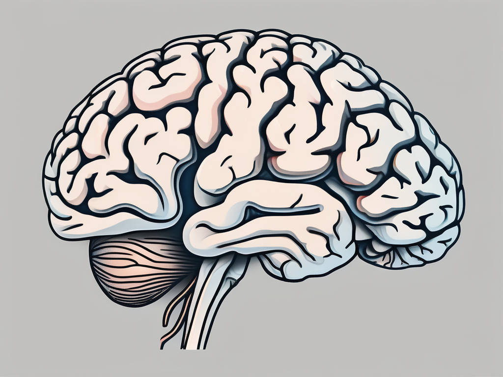
The trochlear nerve, also known as the fourth cranial nerve, is an important component of the central nervous system. It plays a crucial role in the coordination of eye movement and enables us to focus our vision on different objects. Understanding the pathway and function of the trochlear nerve is essential for comprehending the complexity of our visual system.
Understanding the Trochlear Nerve
The trochlear nerve, also known as cranial nerve IV, is a fascinating component of the nervous system. It originates from the midbrain, specifically the dorsal aspect of the brainstem. Unlike the other cranial nerves, the trochlear nerve emerges from the posterior aspect of the brainstem, giving it a unique position and pathway.
Let’s delve deeper into the anatomy and function of the trochlear nerve to gain a comprehensive understanding of its role in the intricate workings of the human body.
Anatomy of the Trochlear Nerve
The trochlear nerve consists of motor fibers that innervate the superior oblique muscle of the eye. It originates from the trochlear nucleus, which is located within the midbrain. This nucleus serves as the command center for the trochlear nerve, orchestrating the precise movements of the superior oblique muscle.
From its origin, the fibers of the trochlear nerve exit the brainstem dorsally and cross the contralateral midbrain. This crossing ensures that the appropriate signals are sent to the correct eye, allowing for coordinated movements. After this crucial crossing, the trochlear nerve proceeds to pass through the cavernous sinus, a complex network of veins and nerves.
As the trochlear nerve traverses the cavernous sinus, it embarks on a remarkable journey towards the eye. It enters the orbit, the bony cavity that houses the eyeball, via the superior orbital fissure. This narrow passageway serves as the gateway for the trochlear nerve, guiding it towards its ultimate destination.
Within the orbit, the trochlear nerve takes a unique path. It wraps around the annulus of Zinn, a fibrous ring-like structure, before reaching the superior oblique muscle. This intricate anatomical pathway ensures that the trochlear nerve can precisely innervate the superior oblique muscle, enabling smooth and coordinated eye movements.
Function of the Trochlear Nerve
The trochlear nerve plays a crucial role in controlling the superior oblique muscle of the eye. This muscle is responsible for various eye movements, particularly depression (downward movement) and intorsion (inward rotation). Activation of the trochlear nerve triggers the contraction of the superior oblique muscle, facilitating these essential eye movements.
To understand how the trochlear nerve achieves such precise control over eye movements, we must explore the unique pulley system formed by the trochlea. The trochlea is a fibrocartilaginous structure located anterior to the superior oblique muscle. It acts as a guide, ensuring that the trochlear nerve fibers are properly directed, allowing for accurate and coordinated eye movements.
Disruption or damage to the trochlear nerve can lead to various eye movement disorders. These conditions can manifest as double vision, difficulty in looking downward or inward, or an inability to align the eyes with a visual target. Understanding the intricate workings of the trochlear nerve is crucial in diagnosing and treating such disorders, enabling patients to regain optimal eye function.
In conclusion, the trochlear nerve is a remarkable component of the nervous system, responsible for controlling the superior oblique muscle and facilitating precise eye movements. Its unique anatomical pathway and intricate pulley system highlight the intricacies of the human body’s design. By understanding the trochlear nerve’s anatomy and function, we can gain valuable insights into the complexities of the visual system and the importance of this cranial nerve in maintaining optimal eye coordination.
The Pathway of the Trochlear Nerve
The journey of the trochlear nerve begins within the midbrain, where its nucleus is situated. From there, the trochlear nerve fibers decussate, meaning they cross over to the opposite side of the brainstem. This unique feature allows each eye to receive innervation from the contralateral trochlear nucleus.
As the trochlear nerve fibers decussate, they form a compact bundle that traverses through the tegmentum, a region of the midbrain involved in various motor and sensory functions. This pathway is meticulously organized, ensuring precise coordination of eye movements.
Upon crossing over, the trochlear nerve fibers continue their journey through the brainstem, passing through a series of intricate structures. They navigate through the red nucleus, a structure involved in motor coordination, and the substantia nigra, which plays a crucial role in movement control and reward processing.
Origin of the Trochlear Nerve
The trochlear nerve originates from the trochlear nucleus, which is located dorsally within the midbrain. This nucleus consists of a group of neurons that are responsible for controlling the superior oblique muscle of the eye. The axons of the trochlear nucleus converge to form the trochlear nerve, which ultimately exits the brainstem from its dorsal aspect.
As the trochlear nerve emerges from the brainstem, it is surrounded by protective layers of connective tissue, ensuring its integrity and proper function. These layers provide a supportive environment for the trochlear nerve as it embarks on its intricate journey.
Course of the Trochlear Nerve
After crossing over to the contralateral side of the brainstem, the trochlear nerve fibers continue their journey through the cavernous sinus, a complex venous structure located within the skull. This sinus houses a network of blood vessels, cranial nerves, and connective tissue, creating a dynamic environment for the trochlear nerve.
As the trochlear nerve passes through the lateral wall of the cavernous sinus, it interacts with various structures, including the oculomotor nerve, abducens nerve, and the internal carotid artery. These interactions contribute to the intricate network of neural and vascular pathways within the skull.
Upon exiting the cavernous sinus, the trochlear nerve enters the superior orbital fissure, a narrow opening located in the sphenoid bone. This fissure serves as a gateway to the orbit, allowing the trochlear nerve to reach its final destination.
Once inside the orbit, the trochlear nerve continues its course, innervating the superior oblique muscle of the eye. This muscle plays a crucial role in eye movements, allowing for vertical and rotational motions.
The pathway of the trochlear nerve is a remarkable example of the intricate connections and precise coordination within the nervous system. Its journey through the midbrain, brainstem, cavernous sinus, and orbit highlights the complexity and elegance of the human anatomy.
The Trochlear Nerve and the Brainstem
The trochlear nerve’s close association with the brainstem is fundamental in understanding its entry point and collaboration with this vital structure.
The brainstem, located at the base of the brain, is a crucial component of the central nervous system. It serves as a bridge between the spinal cord and the higher brain regions, playing a vital role in regulating essential bodily functions such as breathing, heart rate, and digestion. Additionally, the brainstem is responsible for relaying sensory and motor information between the brain and the rest of the body.
Entry Point into the Brainstem
The trochlear nerve enters the brainstem at the level of the inferior colliculus, which is a part of the midbrain’s auditory pathway. This specific entry point is strategically positioned to ensure proper integration of visual and auditory information within the brainstem.
The inferior colliculus, a structure within the midbrain, is primarily involved in processing auditory stimuli. It receives signals from the ears and relays them to higher brain regions responsible for sound perception and localization. The trochlear nerve’s entry point in close proximity to the inferior colliculus allows for efficient communication between visual and auditory processing centers, facilitating coordinated responses to sensory stimuli.
Role of the Brainstem in Trochlear Nerve Function
The brainstem plays a pivotal role in coordinating eye movements through the integration of various sensory inputs. It receives information from the eyes, inner ear, and other sensory organs, allowing for precise control of eye position and movement.
Within the brainstem, the trochlear nerve interacts with other cranial nerves and neural pathways involved in eye movement control. This collaboration ensures that visual information is accurately processed and translated into appropriate motor commands, enabling the coordination of eye movements with head and body orientation. Such coordination is essential for efficient visual tracking and stabilization, allowing us to smoothly follow moving objects and maintain a clear and stable visual field.
Furthermore, the brainstem’s involvement in trochlear nerve function extends beyond eye movement coordination. It also contributes to the regulation of pupillary size and reflexes, as well as the integration of visual information with other sensory modalities, such as touch and proprioception. This integration enables us to perceive and interact with our environment effectively.
In conclusion, the trochlear nerve’s close association with the brainstem is not only crucial for its entry point into the central nervous system but also for its collaboration with this vital structure. The brainstem’s role in coordinating eye movements and integrating sensory inputs ensures the precise control and efficient functioning of the trochlear nerve, ultimately contributing to our visual perception and interaction with the world around us.
Disorders Related to the Trochlear Nerve
While the trochlear nerve is a sturdy and efficient structure, certain disorders can affect its function and lead to eye movement abnormalities.
The trochlear nerve, also known as the fourth cranial nerve, plays a vital role in controlling the movement of the superior oblique muscle of the eye. This muscle is responsible for rotating the eye downward and inward. Any disruption in the functioning of the trochlear nerve can result in various symptoms and visual disturbances.
Symptoms of Trochlear Nerve Damage
Trochlear nerve damage can manifest as diplopia (double vision), especially when looking downward or inward. This occurs because the affected eye is unable to properly align with the other eye, leading to overlapping images. Patients may also experience difficulty in coordinating eye movements, leading to impaired depth perception and visual tracking.
Individuals with trochlear nerve damage may find it challenging to perform tasks that require precise eye movements, such as reading, driving, or playing sports. The double vision can be particularly bothersome and may cause significant discomfort and visual confusion.
Treatment Options for Trochlear Nerve Disorders
Treatment for trochlear nerve disorders depends on the underlying cause. It is crucial to consult a healthcare professional, such as a neurologist or ophthalmologist, who can accurately diagnose the condition and recommend appropriate management strategies.
In some cases, trochlear nerve damage may be a result of trauma or injury. In such instances, the primary focus of treatment is to address the underlying injury and promote healing. This may involve the use of medications to reduce inflammation, pain, and swelling. Physical therapy exercises may also be recommended to improve eye muscle coordination and strengthen the affected muscles.
For individuals with trochlear nerve disorders caused by underlying medical conditions, such as tumors or infections, a more comprehensive treatment approach may be necessary. This may involve a combination of medication, surgery, and ongoing monitoring to manage the condition effectively.
In severe cases where conservative treatment options fail to provide relief, surgical intervention may be considered. Surgical procedures aimed at repairing or repositioning the affected muscle or addressing any structural abnormalities may be performed by a skilled ophthalmologist or neurosurgeon.
It is important to note that the prognosis for individuals with trochlear nerve disorders can vary depending on the specific cause and severity of the condition. Early diagnosis and prompt treatment can significantly improve outcomes and help individuals regain normal eye function.
The Importance of the Trochlear Nerve in Vision
The trochlear nerve’s significance in vision cannot be overstated, as it plays a crucial role in the coordination of eye movements and ensures our ability to focus on visual targets.
The trochlear nerve, also known as the fourth cranial nerve, is one of the twelve cranial nerves that originate from the brainstem. It is the smallest cranial nerve and has a unique pathway compared to the other cranial nerves. It emerges from the dorsal aspect of the brainstem, just below the inferior colliculus, and then crosses over to the opposite side before innervating the superior oblique muscle of the eye.
The Trochlear Nerve and Eye Movement
Eye movements are essential for various visual tasks, such as following moving objects, scanning the environment, and maintaining stable gaze. The trochlear nerve’s precise control over the superior oblique muscle allows for the accurate movement of the eyes, facilitating efficient vision.
The superior oblique muscle is responsible for rotating the eye downward and outward. When the trochlear nerve is functioning properly, it ensures that the superior oblique muscle contracts appropriately, allowing the eye to move smoothly and accurately. This coordinated movement of the eyes is crucial for depth perception, as it helps align the visual axes of both eyes, enabling binocular vision and the perception of three-dimensional space.
Additionally, the trochlear nerve also plays a role in the vertical movement of the eye. It helps in the downward rotation of the eye, allowing us to look down and focus on objects located below our line of sight. This movement is particularly important for activities such as reading, writing, and even walking, as it helps us navigate our surroundings effectively.
Impact of Trochlear Nerve Damage on Vision
Trochlear nerve damage can have a profound impact on vision, leading to difficulties in eye movement control and coordination. This can result in reduced visual acuity, impaired depth perception, and challenges in daily activities that rely heavily on accurate eye movements.
Damage to the trochlear nerve can occur due to various reasons, including trauma, infections, tumors, or vascular disorders. When the trochlear nerve is affected, it can lead to a condition known as trochlear nerve palsy. Trochlear nerve palsy is characterized by a weakness or paralysis of the superior oblique muscle, causing the affected eye to deviate upward and inward. This misalignment of the eyes can result in double vision (diplopia) and difficulties in performing tasks that require precise eye movements.
In conclusion, understanding the entry point of the trochlear nerve into the brainstem provides valuable insight into its intricate pathway and crucial role in eye movement coordination. Disorders related to the trochlear nerve can significantly impact vision, emphasizing the importance of seeking professional medical advice and appropriate treatment options. The trochlear nerve stands as a testament to the remarkable complexity and precision of our visual system, showcasing the marvels of human anatomy and function.
