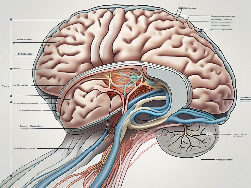
The trochlear nerve, also known as the fourth cranial nerve, is an important component of the human nervous system. It plays a vital role in facilitating eye movement by innervating specific muscles. Understanding the intricacies of this nerve and its associated muscles can provide invaluable insight into the functioning of the human body.
Understanding the Trochlear Nerve
The trochlear nerve is one of the twelve cranial nerves that emerge directly from the brain. Its unique location distinguishes it from its counterparts, as it is the only cranial nerve to emerge from the dorsal surface of the brainstem. This nerve is responsible for the innervation of a single muscle, the superior oblique muscle, which plays a crucial role in eye movement.
The trochlear nerve, also known as the fourth cranial nerve or CN IV, is a fascinating component of the nervous system. Its intricate anatomy and specialized function make it an essential player in the complex orchestra of eye movements.
Anatomy of the Trochlear Nerve
The trochlear nerve contains both motor and proprioceptive fibers, enabling it to transmit signals from the brain to the superior oblique muscle. Beginning at its origin in the midbrain, the nerve traverses a complex pathway to reach its target muscle. This intricate path involves avoiding structures such as the cavernous sinus and the superior orbital fissure. Despite its relatively small size compared to other cranial nerves, the trochlear nerve’s precise anatomy is essential for its proper functioning.
The trochlear nerve is the longest intracranial nerve, measuring approximately 45-55 mm in length. It emerges from the dorsal aspect of the brainstem, specifically the midbrain, near the inferior colliculus. From its origin, the nerve takes a unique course, looping around the brainstem before decussating (crossing over) at the level of the superior medullary velum. This decussation is a crucial step in the trochlear nerve’s pathway, allowing it to innervate the contralateral superior oblique muscle.
After crossing over, the trochlear nerve continues its journey through the subarachnoid space, passing between the posterior cerebral artery and the superior cerebellar artery. It then enters the cavernous sinus, a venous structure located on the lateral aspect of the sella turcica. Within the cavernous sinus, the trochlear nerve travels alongside other cranial nerves, including the oculomotor nerve (CN III), the ophthalmic division of the trigeminal nerve (CN V1), and the abducens nerve (CN VI).
Leaving the cavernous sinus, the trochlear nerve enters the orbit through the superior orbital fissure. This narrow opening provides a pathway for the nerve to reach its target muscle, the superior oblique. The superior oblique muscle, along with the trochlear nerve, forms a unique anatomical relationship. The muscle passes through a fibrocartilaginous loop called the trochlea, which acts as a pulley, changing the direction of the muscle’s pull. This pulley-like structure is what gives the trochlear nerve its name.
Function of the Trochlear Nerve
The primary function of the trochlear nerve is to control the superior oblique muscle, one of the six extraocular muscles responsible for eye movement. The superior oblique muscle has a unique pulley-like structure known as the trochlea, which the trochlear nerve is named after. When the trochlear nerve is stimulated, it causes contraction of the superior oblique muscle, resulting in specific eye movements. These movements allow for the rotation, elevation, and depression of the eye, contributing to visual stability.
Eye movements are complex and precisely coordinated actions that involve multiple cranial nerves, including the trochlear nerve. The trochlear nerve’s role in eye movement is particularly intriguing due to its unique anatomical pathway and the specialized function of the superior oblique muscle.
When the trochlear nerve is activated, it initiates a series of events that lead to the contraction of the superior oblique muscle. This contraction causes the eye to rotate inward (intorsion), move downward (depression), and tilt away from the midline (abduction). These movements are crucial for maintaining proper alignment of the eyes and ensuring clear vision.
In addition to its motor function, the trochlear nerve also contains proprioceptive fibers. These fibers provide sensory feedback from the superior oblique muscle to the brain, allowing for precise coordination of eye movements. This proprioceptive information helps the brain to continuously monitor and adjust the position of the eyes, ensuring optimal visual tracking and stability.
Understanding the trochlear nerve’s anatomy and function is essential for diagnosing and treating disorders that affect eye movements. Damage or dysfunction of the trochlear nerve can lead to a condition known as trochlear nerve palsy, characterized by weakness or paralysis of the superior oblique muscle. This can result in a variety of symptoms, including double vision, difficulty with downward gaze, and abnormal head posture.
In conclusion, the trochlear nerve is a remarkable cranial nerve that plays a crucial role in eye movement. Its unique anatomy and specialized function make it an intriguing component of the nervous system. By understanding the intricacies of the trochlear nerve, healthcare professionals can better diagnose and manage conditions that affect eye movements, ultimately improving the quality of life for individuals with these disorders.
Muscles Innervated by the Trochlear Nerve
In addition to the superior oblique muscle, the trochlear nerve indirectly affects other muscles involved in eye movements. The coordinated functioning of these muscles is essential for maintaining ocular alignment and allowing for smooth eye movements. The following muscles are innervated either directly or indirectly by the trochlear nerve:
Superior Oblique Muscle
The superior oblique muscle is the primary muscle innervated by the trochlear nerve. Located at the back of the eye, it arises from the upper surface of the eye socket and passes through the trochlea. When the trochlear nerve contracts the superior oblique muscle, it assists in depression, abduction, and internal rotation of the eye, allowing for precise eye movements.
But what exactly does it mean for the superior oblique muscle to assist in depression, abduction, and internal rotation of the eye? Let’s break it down. Depression refers to the downward movement of the eye, which is crucial for looking down or shifting the gaze towards the floor. Abduction, on the other hand, involves moving the eye away from the midline of the face, allowing for lateral movements. Lastly, internal rotation refers to the inward rotation of the eye, which is necessary for certain visual tasks like reading or focusing on nearby objects.
Role of the Trochlear Nerve in Eye Movement
Beyond the superior oblique muscle, the trochlear nerve indirectly affects other extraocular muscles responsible for eye movement. These muscles include the inferior oblique, lateral rectus, and medial rectus muscles, all of which collaborate to produce coordinated eye movements. The trochlear nerve’s role in innervating the superior oblique muscle, combined with its interactions with other cranial nerves, ensures synchronized eye movements and precise visual tracking.
Let’s delve deeper into the role of the trochlear nerve in eye movement. The inferior oblique muscle, innervated by the oculomotor nerve, works in conjunction with the superior oblique muscle to produce vertical eye movements. The lateral rectus muscle, innervated by the abducens nerve, is responsible for outward eye movements, allowing the eye to look towards the side. On the other hand, the medial rectus muscle, also innervated by the oculomotor nerve, facilitates inward eye movements, enabling the eye to look towards the midline.
All of these muscles, including the superior oblique muscle, play a vital role in maintaining ocular alignment and allowing for smooth eye movements. Without the precise coordination of these muscles, our ability to track moving objects, read, and maintain binocular vision would be compromised.
Disorders Related to the Trochlear Nerve
Like any other cranial nerve, the trochlear nerve is susceptible to various disorders that can affect its functioning. One such disorder is trochlear nerve palsy, a condition characterized by weakness or paralysis of the superior oblique muscle. Trochlear nerve palsy can lead to vertical diplopia (double vision), an inability to align the eyes correctly, and difficulties with tasks such as reading or driving.
Trochlear nerve palsy can occur due to trauma, infection, or underlying medical conditions such as diabetes or elevated intracranial pressure. The trochlear nerve, also known as the fourth cranial nerve, is responsible for the movement of the superior oblique muscle, which controls the downward and inward rotation of the eye. When the trochlear nerve is affected, it can disrupt the normal coordination between the eyes, leading to vision disturbances.
Symptoms of trochlear nerve palsy may present as a downward head tilt or a compensatory head turn to minimize diplopia. Individuals experiencing any vision-related difficulties or symptoms should consult with a healthcare professional for a proper diagnosis and treatment options. Early intervention is crucial to prevent further complications and improve the quality of life for those affected.
Trochlear Nerve Palsy
Trochlear nerve palsy is a relatively rare condition, accounting for only a small percentage of all cranial nerve disorders. It can occur at any age, but it is more commonly seen in adults. The exact cause of trochlear nerve palsy can vary, and in some cases, the underlying cause may remain unknown.
Diagnosing trochlear nerve palsy requires a thorough evaluation by a healthcare professional. This evaluation may include a comprehensive medical history, physical examination, and specialized tests to assess eye movements and muscle function. The healthcare professional will also inquire about any recent trauma or infections that may have contributed to the development of trochlear nerve palsy.
Treatment options for trochlear nerve palsy depend on the underlying cause and severity of symptoms. In mild cases, conservative approaches may be employed to manage the condition. These approaches may include the use of prism glasses, which help to correct the alignment of the eyes and reduce double vision. Eye exercises may also be recommended to strengthen the muscles and improve coordination.
In more severe cases of trochlear nerve palsy, surgical interventions may be necessary. These interventions aim to correct muscle alignment or relieve pressure on the nerve. The specific surgical procedure will depend on the individual’s condition and the recommendations of the healthcare professional.
Diagnosis and Treatment of Trochlear Nerve Disorders
Diagnosing trochlear nerve disorders requires a comprehensive evaluation by a qualified healthcare professional. This evaluation may include a thorough medical history, physical examination, and specialized tests to assess eye movements and muscle function. The healthcare professional will also consider other potential causes of the symptoms, such as underlying medical conditions or previous trauma.
Once a diagnosis is made, the healthcare professional will discuss the treatment options with the individual. Treatment for trochlear nerve disorders depends on the underlying cause and severity of symptoms. In some cases, the disorder may resolve on its own with time and conservative management. However, in other cases, more targeted interventions may be necessary.
Conservative approaches, such as prism glasses or eye exercises, may be employed to manage mild cases of trochlear nerve disorders. Prism glasses help to correct the alignment of the eyes and reduce double vision, while eye exercises can strengthen the muscles and improve coordination.
In more severe cases, surgical interventions may be required. These interventions aim to correct muscle alignment or relieve pressure on the nerve. The specific surgical procedure will depend on the individual’s condition and the recommendations of the healthcare professional.
It is important for individuals experiencing any vision-related difficulties or symptoms to consult with a healthcare professional for a proper diagnosis and treatment plan. Early intervention can help prevent further complications and improve the overall prognosis for those affected by trochlear nerve disorders.
The Trochlear Nerve in the Nervous System
The trochlear nerve, also known as cranial nerve IV, is a vital component of the nervous system. While its primary role is associated with eye movement, it also maintains a close relationship with other cranial nerves and contributes to overall nervous system functioning.
The trochlear nerve emerges from the dorsal aspect of the midbrain, specifically from the trochlear nucleus. It is the smallest cranial nerve in terms of the number of axons it contains. These axons exit the brainstem and travel along a complex pathway, ultimately reaching the superior oblique muscle of the eye.
Connection with Other Cranial Nerves
The trochlear nerve works in harmony with the other cranial nerves to ensure coordinated eye movements and provide sensory input from the ocular muscles. Particularly, its interactions with the oculomotor nerve, which innervates the majority of extraocular muscles, help facilitate the precise functionality of the eye. The trochlear nerve and the oculomotor nerve work together to control the movements of the eye in different directions, allowing for smooth and accurate visual tracking.
In addition to its connection with the oculomotor nerve, the trochlear nerve also interacts with the abducens nerve. The abducens nerve is responsible for the lateral movement of the eye, while the trochlear nerve primarily controls the downward and inward rotation of the eye. These coordinated movements ensure that the eyes work together to focus on objects of interest and maintain binocular vision.
Any disruption in these connections can lead to impaired eye movements and subsequent visual disturbances. Conditions such as trochlear nerve palsy, where the trochlear nerve is damaged or compressed, can result in double vision, difficulty in looking downward, and an abnormal head tilt to compensate for the impaired eye movement.
Importance of the Trochlear Nerve in Overall Health
Although the trochlear nerve’s primary focus is eye movement, its influence extends beyond ocular functionality. By contributing to the complex network of cranial nerves, it plays a crucial role in maintaining overall health and well-being. The trochlear nerve’s involvement in eye movements and ocular alignment ensures optimal visual capabilities, which are integral to various daily activities and quality of life.
In addition to its role in eye movement, the trochlear nerve also provides sensory feedback from the ocular muscles to the brain. This feedback loop allows for constant monitoring and adjustment of eye position, ensuring that the visual system remains accurate and responsive.
Furthermore, the trochlear nerve’s connections with other cranial nerves contribute to the regulation of other bodily functions. For example, the trochlear nerve has been found to have connections with the trigeminal nerve, which is responsible for sensation in the face and control of the muscles involved in chewing. This interplay between cranial nerves highlights the intricate nature of the nervous system and its impact on various physiological processes.
In conclusion, the trochlear nerve is a crucial component of the nervous system, with its primary role in eye movement and ocular alignment. Its interactions with other cranial nerves ensure coordinated eye movements and contribute to overall health and well-being. Understanding the intricate connections and functions of the trochlear nerve provides valuable insights into the complexity of the human body and its remarkable ability to perceive and interact with the world.
In Conclusion
The trochlear nerve’s innervation of the superior oblique muscle is crucial for facilitating precise eye movements and maintaining proper ocular alignment. Understanding the anatomy, function, and associated disorders of this important cranial nerve provides valuable insights into the intricate workings of the human body. If you experience any vision-related symptoms or concerns, it is always recommended to consult with a healthcare professional for an accurate diagnosis and appropriate management.
