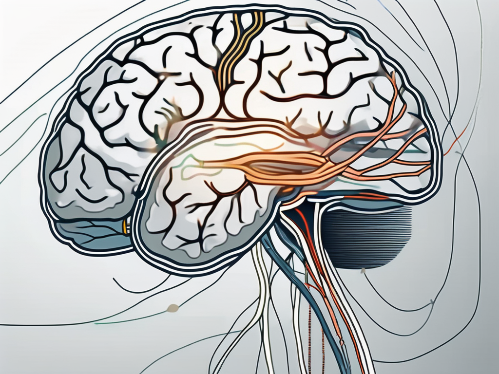
In the realm of medical conditions affecting the eyes, trochlear nerve palsy is a complex and noteworthy condition that requires careful consideration. To fully comprehend its impact, it is crucial to understand the intricacies of trochlear nerve palsy, the role it plays in vision, and the potential causes behind it.
Understanding Trochlear Nerve Palsy
First and foremost, trochlear nerve palsy refers to the dysfunction or impairment of the fourth cranial nerve, known as the trochlear nerve or cranial nerve IV. The trochlear nerve is responsible for controlling the superior oblique muscle, which aids in the movement of the eyes. When this nerve is affected, it can significantly disrupt the normal functioning of the eyes.
Trochlear nerve palsy is a condition that can have a profound impact on a person’s vision and overall quality of life. Understanding the definition, function, and common causes of this condition is crucial in order to provide appropriate care and support to those affected.
Definition and Function of the Trochlear Nerve
The trochlear nerve, as mentioned earlier, is the fourth cranial nerve. It originates in the trochlear nucleus, which is located in the midbrain. The primary function of the trochlear nerve is to innervate the superior oblique muscle. This muscle assists in the downward and inward rotation of the eye, allowing for proper eye movements and alignment.
When the trochlear nerve is functioning properly, it works in harmony with the other cranial nerves to ensure smooth and coordinated eye movements. However, when there is dysfunction or impairment of the trochlear nerve, it can lead to a range of symptoms and visual disturbances.
Individuals with trochlear nerve palsy may experience double vision, difficulty with upward or downward gaze, or a tilted or rotated appearance of objects. These symptoms can significantly impact daily activities such as reading, driving, and even walking, as they rely heavily on accurate eye movements.
Common Causes of Trochlear Nerve Palsy
Trochlear nerve palsy can occur as a result of various underlying causes. It can be congenital, meaning present at birth, or acquired later in life. Common causes of trochlear nerve palsy include head trauma, infections, tumors, vascular abnormalities, and certain systemic conditions such as diabetes or multiple sclerosis.
Head trauma, such as a severe blow to the head or a skull fracture, can damage the trochlear nerve and lead to palsy. Infections, such as meningitis or encephalitis, can also affect the nerve and result in palsy. Tumors, both benign and malignant, can compress or infiltrate the trochlear nerve, causing dysfunction. Vascular abnormalities, such as aneurysms or arteriovenous malformations, can disrupt the blood supply to the nerve and lead to palsy.
Furthermore, certain systemic conditions like diabetes or multiple sclerosis can also contribute to trochlear nerve palsy. Diabetes, for example, can cause damage to the nerves throughout the body, including the trochlear nerve. Multiple sclerosis, an autoimmune disease that affects the central nervous system, can lead to inflammation and demyelination of the nerve, resulting in palsy.
It is important to note that the underlying cause of trochlear nerve palsy may vary from person to person. A thorough medical evaluation and diagnostic testing are necessary to determine the specific cause and guide appropriate treatment options.
The Impact of Right Trochlear Nerve Palsy
When trochlear nerve palsy affects the right eye, it can provoke a range of symptoms and significantly impact a person’s vision and quality of life. Recognizing these signs is crucial for early detection and appropriate management of the condition.
Symptoms and Signs to Look Out For
Individuals with right trochlear nerve palsy often experience symptoms such as double vision, particularly when looking downward or inward. They may also exhibit an abnormal head posture, tilting their head away from the affected eye to minimize diplopia or double vision. Other symptoms may include eye misalignment, difficulty focusing, and reduced depth perception.
How Right Trochlear Nerve Palsy Affects Vision
The impact of right trochlear nerve palsy on vision is profound. Due to the weakened function of the superior oblique muscle, the affected eye may have limited ability to move downward and inward. The resultant misalignment of the eyes can lead to binocular vision problems, which can potentially affect daily activities such as reading, driving, or even simple tasks such as walking down stairs.
Furthermore, the reduced depth perception caused by right trochlear nerve palsy can have significant consequences on a person’s spatial awareness and coordination. Tasks that require judging distances accurately, such as catching a ball or pouring a glass of water, may become challenging and frustrating.
Moreover, the impact of right trochlear nerve palsy extends beyond the physical limitations it imposes. The psychological effects of living with a visual impairment can be profound. Individuals may experience feelings of frustration, helplessness, and a loss of independence. Simple activities that were once taken for granted, such as reading a book or watching a movie, may become arduous tasks that require extra effort and concentration.
In addition to the physical and psychological effects, right trochlear nerve palsy can also have social implications. The visible signs of eye misalignment and abnormal head posture can attract unwanted attention and lead to self-consciousness. Individuals may feel uncomfortable in social situations and may avoid eye contact, leading to potential difficulties in communication and social interactions.
It is important to note that the impact of right trochlear nerve palsy can vary from person to person. Some individuals may adapt and find strategies to cope with the visual challenges, while others may require more extensive interventions such as surgery or vision therapy to improve their quality of life.
Diagnosis of Right Trochlear Nerve Palsy
Accurate diagnosis of right trochlear nerve palsy is crucial to guide appropriate treatment strategies. Healthcare professionals employ various methods to determine the underlying cause and severity of the condition.
Medical History and Physical Examination
During the diagnostic process, doctors will take a detailed medical history and conduct a thorough physical examination. This helps identify any potential underlying conditions or triggering factors that may have led to trochlear nerve palsy.
When obtaining the medical history, the doctor will inquire about any recent head injuries, infections, or surgeries that the patient may have had. They will also ask about any symptoms the patient is experiencing, such as double vision, eye pain, or difficulty moving the affected eye.
The physical examination will involve a comprehensive assessment of the patient’s eye movements, visual acuity, and pupillary reflexes. The doctor will carefully observe the alignment and coordination of the eyes, looking for any signs of misalignment or abnormal eye movements.
In addition, the doctor may perform specific tests to evaluate the function of the trochlear nerve. These tests may include the Parks-Bielschowsky three-step test, which assesses the vertical and torsional movements of the affected eye.
Imaging and Other Diagnostic Tests
In addition to a physical examination, medical imaging tests such as magnetic resonance imaging (MRI) or computed tomography (CT) scans may be recommended. These diagnostic tools allow healthcare providers to visualize any structural abnormalities or lesions that could be affecting the trochlear nerve.
An MRI scan uses a powerful magnetic field and radio waves to create detailed images of the brain and cranial nerves. This imaging technique can help identify any tumors, vascular malformations, or other lesions that may be compressing or damaging the trochlear nerve.
A CT scan, on the other hand, uses X-rays and a computer to generate cross-sectional images of the head and brain. This imaging modality can provide valuable information about the bony structures surrounding the trochlear nerve, such as fractures or abnormalities.
In some cases, additional diagnostic tests may be performed to further evaluate the function of the trochlear nerve. These tests may include electrodiagnostic studies, such as electromyography (EMG) or nerve conduction studies (NCS), which measure the electrical activity and conduction velocity of the nerve.
Furthermore, blood tests may be ordered to check for any underlying systemic conditions or autoimmune disorders that could be contributing to the trochlear nerve palsy.
Treatment Options for Right Trochlear Nerve Palsy
Right trochlear nerve palsy is a condition that requires careful consideration of various treatment options. The severity of the condition, underlying causes, and individual patient characteristics all play a role in determining the most suitable approach.
Non-surgical treatments are often the first line of management for right trochlear nerve palsy. These treatments aim to alleviate symptoms and improve eye function without the need for invasive procedures. One such non-surgical option is the use of prism glasses, which can correct double vision and provide relief to patients. Another approach is eye patching, which can help alleviate symptoms and promote healing. Additionally, vision therapies focused on improving eye alignment and coordination may be recommended to enhance the patient’s visual capabilities.
It is important to note that each case of right trochlear nerve palsy is unique, and consulting with a healthcare professional is crucial in determining the most appropriate treatment plan. A thorough evaluation of the patient’s condition, medical history, and individual needs will help guide the treatment decisions.
In some cases, non-surgical approaches may prove ineffective or the palsy may be severe, necessitating surgical interventions. Surgical options can help correct ocular misalignment and enhance eye movements, leading to improved visual function. One such surgical procedure is trochleoplasty, which involves reshaping the trochlear groove to improve the movement of the affected eye. Eye muscle surgery is another surgical intervention that may be considered, aiming to adjust the tension and alignment of the eye muscles to restore proper eye movement.
However, it is crucial to undergo a thorough evaluation and consult with an ophthalmologist or other medical experts to assess the suitability of surgical options. The decision to pursue surgery should be based on a comprehensive understanding of the patient’s condition, potential risks and benefits, and the likelihood of achieving the desired outcomes.
Living with Right Trochlear Nerve Palsy
Adjusting to life with right trochlear nerve palsy can be challenging, but various coping strategies and lifestyle adjustments can help improve quality of life for those affected.
Right trochlear nerve palsy is a condition that affects the fourth cranial nerve, which controls the movement of the superior oblique muscle of the eye. This muscle is responsible for downward and inward rotation of the eye. When the trochlear nerve is damaged or impaired, it can lead to a range of symptoms, including double vision, difficulty with depth perception, and eye misalignment.
Living with right trochlear nerve palsy requires individuals to adapt to the changes in their vision and find ways to manage the associated symptoms. Coping strategies and lifestyle adjustments can play a crucial role in improving functionality and overall well-being.
Coping Strategies and Lifestyle Adjustments
Individuals living with trochlear nerve palsy can benefit from seeking support and guidance from healthcare professionals specializing in vision therapy, as well as support groups. Vision therapy involves a series of exercises and activities designed to improve eye coordination and strengthen the muscles responsible for eye movement.
In addition to vision therapy, modifying daily routines and workplace environments can help minimize eye strain and optimize vision. This may include adjusting lighting conditions, using specialized eyewear or filters to reduce glare, and taking regular breaks to rest the eyes.
Furthermore, individuals with trochlear nerve palsy may find it helpful to explore assistive devices and technologies that can aid in daily activities. For example, magnifying glasses or screen readers can assist with reading, while adaptive tools like large-print calendars or voice-activated reminders can help with organization and time management.
Long-Term Prognosis and Quality of Life
It is essential to approach right trochlear nerve palsy with a long-term perspective. While the severity of the condition varies among individuals, with appropriate management and adherence to treatment plans, many patients can achieve significant improvements in their symptoms and quality of life.
Regular follow-ups with healthcare providers will allow for close monitoring and adjustments to treatment if necessary. These appointments may include comprehensive eye exams, visual field tests, and evaluations of eye muscle function. By closely monitoring the condition, healthcare professionals can identify any changes or progression of symptoms and make appropriate recommendations for treatment adjustments or interventions.
Additionally, maintaining a healthy lifestyle can contribute to overall well-being and potentially improve the prognosis of trochlear nerve palsy. This includes eating a balanced diet rich in nutrients that support eye health, engaging in regular exercise to promote circulation and reduce the risk of other health conditions, and getting enough sleep to support optimal eye function and recovery.
In conclusion, right trochlear nerve palsy can have a profound impact on an individual’s vision and overall well-being. Recognizing the signs, seeking early diagnosis, and exploring appropriate treatment options are key in managing this condition. With the guidance of medical professionals, individuals can navigate the challenges and embark on a path towards improved eye health and functionality.
