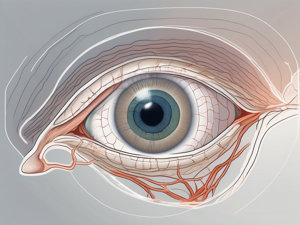
Understanding the anatomy and functionality of the human eye is crucial to comprehend the role of various components in vision. One such important aspect is the trochlear nerve, which controls an extrinsic eye muscle known as the superior oblique muscle. In this article, we will delve into the intricate details of this fascinating connection and explore the potential disorders associated with the trochlear nerve that can impact vision.
Understanding the Anatomy of the Eye
To fully grasp the significance of the trochlear nerve’s role in eye function, we must first understand the complex structure of the eye itself. The eye is composed of multiple components, including the cornea, iris, lens, retina, and optic nerve. These elements work together seamlessly to enable us to perceive the world around us.
Starting with the cornea, it is the transparent front part of the eye that covers the iris, pupil, and anterior chamber. It plays a crucial role in refracting light and focusing it onto the retina. The iris, on the other hand, is the colored part of the eye that controls the size of the pupil, regulating the amount of light entering the eye.
The lens, located behind the iris, further helps in focusing light onto the retina. It changes shape to adjust the eye’s focus, allowing us to see objects at different distances clearly. The retina, a thin layer of tissue at the back of the eye, contains millions of specialized cells called photoreceptors that convert light into electrical signals, which are then transmitted to the brain via the optic nerve.
Speaking of the optic nerve, it is a bundle of more than a million nerve fibers that carry visual information from the retina to the brain. This vital connection allows us to process and interpret what we see, forming our visual perception of the world.
Additionally, the eye is equipped with a set of extrinsic eye muscles, which are responsible for controlling the movement of the eyeball. These muscles, six in total, work in harmony to enable precise eye movements in all directions.
The Role of Extrinsic Eye Muscles
The extrinsic eye muscles play a crucial role in ensuring coordinated eye movements and maintaining visual alignment. These muscles attach to the outer surface of the eye and control its movement in different directions – up, down, left, and right – as well as rotational movements.
Each of these muscles has a specific function. The superior rectus muscle, for example, is responsible for upward eye movement, while the inferior rectus muscle controls downward eye movement. The lateral rectus muscle allows the eye to move outward, while the medial rectus muscle enables inward movement. The superior oblique muscle helps in downward and inward eye movement, while the inferior oblique muscle assists in upward and outward eye movement.
Collectively, these muscles enable us to explore our environment by allowing us to shift our gaze smoothly and accurately. Dysfunction of any of these muscles can lead to various vision-related problems, such as strabismus (crossed eyes) or nystagmus (involuntary eye movement).
The Function of the Trochlear Nerve
In this intricate web of eye muscles, the trochlear nerve holds a significant position. The trochlear nerve, also known as the fourth cranial nerve, is responsible for controlling the superior oblique muscle.
Located in the midbrain, this nerve has the longest intracranial course among all the cranial nerves. Its unique trajectory and connection to the superior oblique muscle make it a critical component in eye movement and coordination.
When the trochlear nerve is functioning properly, it allows the superior oblique muscle to contract and move the eye in a downward and inward direction. This movement is essential for various visual tasks, such as reading, tracking moving objects, and maintaining proper eye alignment.
Any damage or dysfunction of the trochlear nerve can result in a condition known as trochlear nerve palsy, which leads to weakness or paralysis of the superior oblique muscle. This can cause double vision, difficulty in looking downward, and problems with depth perception.
In conclusion, understanding the intricate anatomy of the eye and the role of the trochlear nerve provides us with a deeper appreciation for the complexity and precision involved in our visual perception. The collaboration between the eye’s components and the coordination of the extrinsic eye muscles are essential for our ability to see and navigate the world around us.
The Trochlear Nerve and the Superior Oblique Muscle
Now, let us explore the connection between the trochlear nerve and the superior oblique muscle in further detail to comprehend their interdependence and influence on vision.
The Connection between the Trochlear Nerve and the Superior Oblique Muscle
The trochlear nerve originates from the dorsal midbrain and courses through the skull, eventually meeting the superior oblique muscle. This muscle is responsible for certain eye movements, including depression (looking downwards) and intorsion (rotating the eye towards the nose).
Remarkably, the trochlear nerve is the only cranial nerve that emerges from the back of the brainstem, allowing it to function independently and control the superior oblique muscle’s unique movements.
The trochlear nerve’s journey from the midbrain to the superior oblique muscle involves traversing a complex pathway within the skull. It passes through the superior orbital fissure, a narrow opening located in the sphenoid bone. This intricate route ensures that the trochlear nerve reaches its target muscle with precision and accuracy.
Upon reaching the superior oblique muscle, the trochlear nerve establishes a vital connection that enables the coordination of eye movements. This connection is essential for maintaining proper alignment and stability of the eyes, allowing for smooth and accurate visual tracking.
How the Trochlear Nerve Controls the Superior Oblique Muscle
The trochlear nerve innervates the superior oblique muscle, meaning it supplies the necessary nerve fibers for the muscle’s contraction and control. This union between the trochlear nerve and the superior oblique muscle allows for the desired eye movements to be executed accurately and efficiently.
When the trochlear nerve is functioning optimally, it transmits signals to the superior oblique muscle, regulating its contractions and movements. This precise control ensures that the eye can move in the desired direction, contributing to clear and focused vision.
Moreover, the trochlear nerve’s unique positioning at the back of the brainstem grants it a degree of independence in controlling the superior oblique muscle. This independence allows for fine-tuned adjustments in eye movements, making it possible to navigate complex visual environments with ease.
However, when there is a disruption or damage to the trochlear nerve, it can lead to a range of symptoms and potential vision problems. Conditions such as trochlear nerve palsy can result in weakness or paralysis of the superior oblique muscle, leading to double vision, difficulty in looking downwards, and an altered perception of depth.
Understanding the intricate relationship between the trochlear nerve and the superior oblique muscle is crucial for comprehending the complexities of vision and the mechanisms that govern eye movements. By delving into the details of this connection, we gain a deeper appreciation for the remarkable coordination and precision required for our eyes to function optimally.
Disorders Related to the Trochlear Nerve
The trochlear nerve and the superior oblique muscle work together harmoniously to facilitate proper eye movement and visual function. However, despite their intricate coordination, these structures are susceptible to various disorders that can significantly impact visual function. It is crucial to be aware of these disorders and seek appropriate medical attention if symptoms arise.
One common disorder related to the trochlear nerve is trochlear nerve palsy. This condition occurs when the trochlear nerve is damaged or impaired, leading to difficulties in eye movement and coordination. Trochlear nerve palsy can result from trauma, infections, tumors, or other underlying medical conditions. The severity of the impairment can vary, ranging from mild to severe.
Symptoms of Trochlear Nerve Damage
The symptoms of trochlear nerve damage can vary depending on the severity of the impairment. Common signs may include double vision, difficulty moving the affected eye, tilted head posture to compensate for eye misalignment, and eyestrain. These symptoms can significantly impact an individual’s quality of life, making it challenging to perform daily activities that require clear and coordinated vision.
Experiencing these symptoms should prompt an immediate consultation with an eye care professional to assess the condition and provide appropriate guidance and treatment. Early detection and intervention can help prevent further complications and improve the chances of successful treatment.
Treatment Options for Trochlear Nerve Disorders
As with any medical condition, the treatment options for trochlear nerve disorders will depend on the specific diagnosis and the individual’s unique circumstances. Consulting with a healthcare professional, such as an ophthalmologist or neurologist, is essential to obtain an accurate diagnosis and determine the most suitable treatment plan.
Treatment approaches may include therapeutic exercises to improve eye movements, surgery to correct muscle imbalances, or the use of prism glasses to alleviate symptoms. Therapeutic exercises, such as eye-tracking exercises and eye muscle strengthening exercises, can help improve eye coordination and movement. Surgical intervention may be necessary in cases where muscle imbalances or structural abnormalities are present. Prism glasses, which contain specially designed lenses, can help correct double vision and improve visual alignment.
Each treatment option will be tailored to the individual’s needs and aimed at optimizing visual function. It is important to follow the recommended treatment plan and attend regular follow-up appointments to monitor progress and make any necessary adjustments.
In conclusion, disorders related to the trochlear nerve can significantly impact visual function. Recognizing the symptoms of trochlear nerve damage and seeking appropriate medical attention is crucial for early intervention and successful treatment. With the help of healthcare professionals, individuals with trochlear nerve disorders can receive the necessary care to improve their visual function and overall quality of life.
The Impact of the Trochlear Nerve on Vision
Understanding the significance of the trochlear nerve in eye movement and coordination allows us to grasp its profound impact on vision.
The trochlear nerve, also known as the fourth cranial nerve, is one of the twelve cranial nerves that emerge directly from the brain. It is the smallest cranial nerve and has the longest intracranial course. Despite its small size, the trochlear nerve plays a crucial role in the complex process of eye movement.
The Role of the Trochlear Nerve in Eye Movement
The trochlear nerve plays a critical role in orchestrating the precise movements of the superior oblique muscle, facilitating both downward gaze and inward rotation. These movements are essential for various activities, such as reading, driving, and participating in sports.
Imagine trying to read a book without the ability to move your eyes smoothly and accurately. The trochlear nerve ensures that our eyes can move effortlessly, allowing us to track words on a page and shift our focus from one point to another. Without the trochlear nerve’s coordination, our vision would be severely impaired, making even the simplest tasks a challenge.
How Trochlear Nerve Disorders Affect Vision
When the trochlear nerve is compromised, whether due to injury, inflammation, or other underlying conditions, it can disrupt the delicate balance of eye movements. This disruption often leads to visual disturbances, including double vision, eye misalignment, and decreased depth perception.
Double vision, also known as diplopia, is a common symptom of trochlear nerve disorders. It occurs when the eyes are unable to align properly, causing two images to be seen instead of one. This can make it difficult to focus on objects, read, or perform tasks that require visual accuracy.
Eye misalignment, known as strabismus, is another consequence of trochlear nerve disorders. In this condition, one eye may turn inward or outward, resulting in a lack of coordination between the two eyes. This can lead to a loss of depth perception and difficulties with judging distances accurately.
It is important to emphasize that the effects of trochlear nerve disorders on vision can vary, and proper evaluation from a healthcare professional is crucial to determine the extent of the impairment and implement appropriate management strategies.
Furthermore, trochlear nerve disorders can have a significant impact on an individual’s quality of life. Tasks that were once effortless, such as driving or playing sports, may become challenging or even impossible. The emotional and psychological toll of these visual impairments should not be overlooked, as individuals may experience frustration, anxiety, and a decreased sense of independence.
In conclusion, the trochlear nerve plays a vital role in eye movement and coordination, directly influencing our ability to see the world around us. When this nerve is compromised, whether due to injury or underlying conditions, it can have profound effects on vision. Understanding the impact of trochlear nerve disorders on visual function is crucial for healthcare professionals to provide appropriate care and support to those affected.
Conclusion: The Importance of the Trochlear Nerve in Eye Function
As we conclude our exploration of the trochlear nerve and its association with the superior oblique muscle, we are reminded of its crucial role in maintaining the synchrony of eye movements and preserving our visual experience.
Understanding the intricate connectivity and functionality within the human eye helps us appreciate the complexity of vision and the importance of seeking professional guidance when faced with potential eye-related concerns.
If you experience any visual symptoms or suspect issues with your eye movements, consult with a qualified healthcare professional who can provide an accurate diagnosis and guide you towards the most appropriate course of action. Remember, prioritizing your eye health is essential for enjoying a life of optimal visual function.
