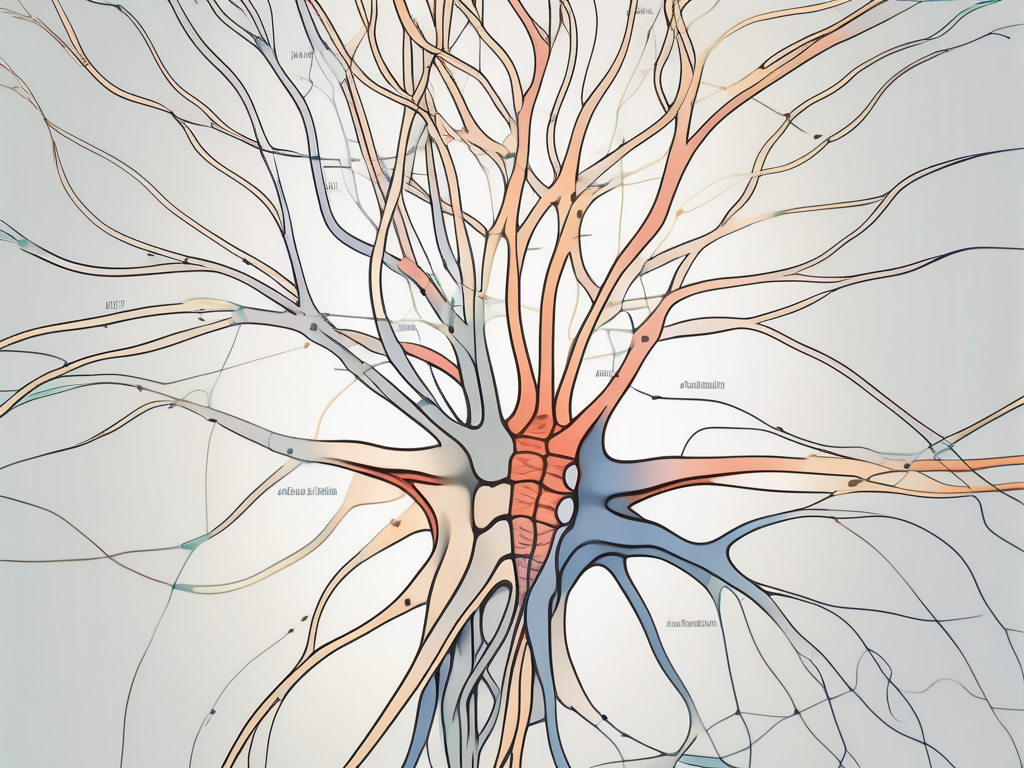
The nervous system is a complex network of cells that plays a crucial role in regulating and coordinating bodily functions. Understanding the structure and function of the nervous system is essential for gaining insights into specific nerves, such as the trochlear nerve, and where their neuron cell bodies are located.
Understanding the Nervous System
The nervous system is a complex and fascinating network that plays a vital role in our bodies. It is divided into two main parts: the central nervous system (CNS) and the peripheral nervous system (PNS). The CNS consists of the brain and spinal cord, which are like the command center of our body, while the PNS includes all the nerve cells outside the CNS, such as those found in the limbs and organs.
Within this intricate system, there are billions of neurons, which are the basic building blocks. Neurons are specialized cells responsible for transmitting electrical signals throughout the body. They are like messengers, delivering important information to different parts of the body.
The Role and Function of the Nervous System
The nervous system serves several crucial functions in the body. One of its primary functions is enabling communication between different parts of the body. This communication allows for coordinated movement and sensory perception. For example, when you touch something hot, the nerves in your skin send a signal to your brain, which then quickly sends a message back to your muscles to move your hand away.
In addition to facilitating communication, the nervous system also regulates vital processes. It plays a key role in controlling automatic functions, such as breathing, heart rate, and digestion. Without the nervous system, these essential processes would not be possible.
Key Components of the Nervous System
As mentioned earlier, the nervous system is made up of neurons, which are the fundamental units of this intricate network. Neurons have a unique structure that allows them to carry out their important functions. Each neuron consists of a cell body, dendrites, and an axon.
The cell body of a neuron houses the nucleus and other essential components necessary for cell function. It is like the control center of the neuron, responsible for coordinating its activities. Dendrites, on the other hand, are branch-like structures that extend from the cell body. They receive signals from other neurons and transmit them to the cell body. Lastly, the axon is a long, slender projection that carries signals away from the cell body to other neurons or target cells.
Together, these components work in harmony to ensure the smooth transmission of electrical signals throughout the nervous system, allowing for efficient communication and coordination within the body.
Introduction to Neurons and their Cell Bodies
Neurons, the building blocks of the nervous system, are fascinating cells that come in various shapes and sizes. While their structures may differ, they generally consist of three main parts: the cell body, dendrites, and axon. Let’s dive deeper into the intricacies of the neuron’s cell body.
The cell body, also known as the soma or perikaryon, is a remarkable hub of activity within the neuron. It is the central region that contains the nucleus, which houses the genetic material responsible for the neuron’s characteristics and functions. Alongside the nucleus, the cell body is home to numerous cellular organelles that work harmoniously to support the neuron’s metabolic functions and maintain its overall health.
The Structure of a Neuron’s Cell Body
When we examine the cell body of a neuron, we discover a world of interconnected structures that contribute to its remarkable capabilities. The nucleus, often likened to the control center of the cell, orchestrates the neuron’s activities by regulating gene expression and synthesizing important proteins.
Surrounding the nucleus, we find mitochondria, the powerhouses of the cell. These tiny organelles generate the energy necessary for the neuron to carry out its functions. They tirelessly produce adenosine triphosphate (ATP), the molecule that fuels the neuron’s activities and ensures its survival.
Furthermore, the endoplasmic reticulum, a complex network of membranes, plays a crucial role in protein synthesis and transport within the cell body. It assists in the folding and modification of proteins, ensuring they are properly shaped and functional.
The Importance of Neuron Cell Bodies
Neuron cell bodies are not mere bystanders in the grand symphony of the nervous system. They play a vital role in the overall function and communication within this intricate network. One of their key responsibilities is the production of neurotransmitters, chemical messengers that enable communication between neurons.
Within the cell body, specialized structures called ribosomes synthesize these neurotransmitters, allowing the neuron to transmit signals to other cells. These chemical messengers are essential for processes such as learning, memory, and motor control.
Moreover, the cell bodies contain the genetic information necessary for the neuron’s survival and proper functioning. The DNA within the nucleus provides the instructions for the synthesis of proteins and other molecules crucial for the neuron’s activities. It ensures that the neuron can adapt to various stimuli and maintain its structural integrity.
In conclusion, the cell body of a neuron is a remarkable and essential component of these extraordinary cells. Its intricate structure and functions contribute to the neuron’s ability to receive, process, and transmit information, ultimately enabling the complex workings of the nervous system.
Overview of the Trochlear Nerve
The trochlear nerve, also known as cranial nerve IV, is one of the twelve cranial nerves that emerge directly from the brain. It is responsible for controlling the superior oblique muscle, which helps with eye movement and coordination.
The Function of the Trochlear Nerve
The trochlear nerve allows for vertical eye movement and helps maintain proper alignment of the eyes during various activities, such as reading, walking, and driving. Without the trochlear nerve’s function, individuals may experience difficulties in moving their eyes up and down, leading to visual disturbances and impaired depth perception.
The Anatomy of the Trochlear Nerve
The trochlear nerve originates in the midbrain, specifically from the trochlear nucleus. As it exits the brainstem, it follows a complex pathway and course towards the superior oblique muscle in each eye. It is the only cranial nerve that emerges from the dorsal (back) side of the brainstem.
Let’s delve deeper into the anatomy of the trochlear nerve. The trochlear nucleus, where the nerve originates, is located in the midbrain, near the cerebral aqueduct. This small nucleus consists of motor neurons that send their axons out to control the superior oblique muscle. The axons of the trochlear nerve exit the midbrain dorsally, meaning they emerge from the backside of the brainstem.
Once the trochlear nerve leaves the midbrain, it takes a unique route through the brainstem. It wraps around the cerebral peduncles, which are bundles of nerve fibers that connect the midbrain to other parts of the brain. The trochlear nerve then crosses the midline and decussates, meaning that the nerve fibers from one side of the brainstem cross over to the opposite side. This decussation allows for coordinated eye movements between the two eyes.
After crossing the midline, the trochlear nerve continues its journey towards the superior oblique muscle. It travels along the inner surface of the skull, passing through the cavernous sinus, a cavity located on each side of the sella turcica, a bony structure in the skull. The cavernous sinus contains various important structures, including blood vessels and other cranial nerves.
As the trochlear nerve approaches the eye, it enters the orbit through the superior orbital fissure, a narrow opening in the skull. Once inside the orbit, the nerve innervates the superior oblique muscle, which is responsible for rotating the eye downward and outward. This movement is crucial for proper eye alignment and coordination.
In summary, the trochlear nerve plays a vital role in eye movement and coordination. Its complex pathway from the midbrain to the superior oblique muscle involves crossing over the midline and passing through important structures in the skull. Understanding the anatomy of the trochlear nerve provides valuable insights into its function and the potential consequences of its dysfunction.
Locating the Neuron Cell Bodies of the Trochlear Nerve
The neuron cell bodies that make up the trochlear nerve are situated within the brainstem.
The Position of the Trochlear Nerve in the Nervous System
The trochlear nerve cell bodies are located in a specific region of the brainstem known as the trochlear nucleus. This nucleus is positioned in the midbrain, a critical part of the central nervous system responsible for relaying sensory and motor information.
The midbrain, also referred to as the mesencephalon, is located between the forebrain and hindbrain. It plays a crucial role in various functions, including visual and auditory processing, motor coordination, and the regulation of sleep and wakefulness.
Within the midbrain, the trochlear nucleus is found dorsally, which means it is situated towards the back of the brainstem. This strategic positioning allows the trochlear nerve to play a vital role in eye movements.
Identifying the Neuron Cell Bodies of the Trochlear Nerve
To precisely identify the neuron cell bodies that make up the trochlear nerve, specialized imaging techniques, such as magnetic resonance imaging (MRI), can be employed. These imaging modalities provide detailed anatomical information, including the precise location of the trochlear nucleus within the brainstem.
MRI utilizes powerful magnets and radio waves to generate detailed images of the internal structures of the body. By applying this technique to the brain, scientists and medical professionals can accurately visualize the trochlear nucleus and its associated neuron cell bodies.
Furthermore, advanced imaging techniques like diffusion tensor imaging (DTI) can provide insights into the connectivity of the trochlear nucleus with other brain regions. DTI measures the diffusion of water molecules in the brain’s white matter tracts, allowing researchers to map the neural pathways associated with the trochlear nerve.
Studying the precise location and connectivity of the trochlear nucleus is crucial for understanding its role in coordinating eye movements. This knowledge can aid in diagnosing and treating various neurological conditions that affect eye movement, such as trochlear nerve palsy or other disorders impacting the midbrain.
Implications of Damage to the Trochlear Nerve
Damage to the trochlear nerve can have significant implications on a person’s visual function and daily activities. The trochlear nerve, also known as the fourth cranial nerve, is responsible for the movement of the superior oblique muscle in the eye. When this nerve is damaged, it can lead to various symptoms and challenges that can greatly impact a person’s quality of life.
Symptoms of Trochlear Nerve Damage
Individuals with trochlear nerve damage may experience diplopia, commonly known as double vision. This occurs particularly when looking downward or to the opposite side. The affected eye may deviate inward or outward, causing a misalignment that results in the perception of two images instead of one. This can be extremely disorienting and make it difficult to focus on objects or perform tasks that require precise visual coordination.
In addition to diplopia, people with trochlear nerve damage may also have difficulty reading. The misalignment of the eyes can make it challenging to track lines of text, leading to blurred or overlapping words. This can significantly impact a person’s ability to engage in activities such as reading books, newspapers, or even digital screens.
Furthermore, judging distances accurately becomes a challenge for individuals with trochlear nerve damage. The misalignment of the eyes can affect depth perception, making it difficult to determine how far away objects are. This can lead to problems with activities that require spatial awareness, such as reaching for objects, catching a ball, or parking a car.
Tasks that involve eye movements, such as walking down stairs or driving in traffic, can also become problematic for those with trochlear nerve damage. The coordination between eye movements and body movements may be disrupted, leading to difficulties in navigating the environment safely and efficiently.
Treatment and Recovery from Trochlear Nerve Damage
Treatment for trochlear nerve damage depends on the underlying cause and severity of the condition. It is essential for individuals experiencing symptoms related to trochlear nerve damage to consult with a healthcare professional, such as a neurologist or ophthalmologist, who can accurately diagnose the condition and develop an appropriate treatment plan.
In some cases, trochlear nerve damage may be caused by trauma, such as head injuries or surgical complications. In these instances, immediate medical attention is crucial to prevent further damage and promote healing. Treatment options may include medications to reduce inflammation, physical therapy to improve eye coordination, or surgical interventions to repair or bypass the damaged nerve.
Recovery from trochlear nerve damage can vary depending on the extent of the injury and the individual’s overall health. Some cases may require long-term management of symptoms, while others may experience partial or complete recovery over time. Rehabilitation exercises and visual therapy can play a significant role in improving eye coordination and minimizing the impact of trochlear nerve damage on daily activities.
It is important for individuals with trochlear nerve damage to receive ongoing support and guidance from healthcare professionals. They can provide valuable information on adaptive strategies, assistive devices, and lifestyle modifications that can help individuals navigate their daily lives more effectively and maintain their independence.
In conclusion, damage to the trochlear nerve can have profound implications on a person’s visual function and overall well-being. Understanding the symptoms and seeking appropriate treatment and support are essential for managing the challenges associated with trochlear nerve damage.
Conclusion: The Significance of Neuron Cell Bodies in the Trochlear Nerve
The neuron cell bodies that make up the trochlear nerve are located within the brainstem, specifically in the trochlear nucleus. These cell bodies are crucial for the proper functioning of the trochlear nerve, which plays a vital role in eye movement and coordination. Understanding the precise location and importance of neuron cell bodies can provide valuable insights into the structure and function of the nervous system and contribute to the development of effective diagnostic and treatment strategies for trochlear nerve-related conditions.
The Impact of Neuron Cell Bodies on Nerve Function
The integrity of neuron cell bodies is crucial for maintaining proper nerve function. Any damage or disruption to these cell bodies can result in impaired signal transmission and, consequently, a range of debilitating symptoms.
Future Research Directions for the Trochlear Nerve
Ongoing research on the trochlear nerve continues to advance our understanding of its function and associated disorders. Studies focusing on neuroregeneration and neuroplasticity may offer promising avenues for enhancing recovery and improving outcomes for individuals with trochlear nerve damage.
In conclusion, the neuron cell bodies that make up the trochlear nerve are found within the brainstem, specifically in the trochlear nucleus. Understanding the anatomical location and significance of these cell bodies contributes to our knowledge of the nervous system and its intricate functioning. As with any medical concerns, it is essential to consult with a healthcare professional for accurate diagnosis and appropriate treatment options.
