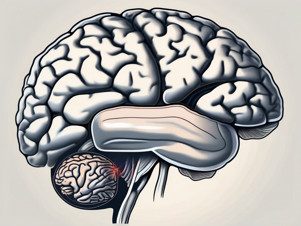
The trochlear nerve is a crucial component of the human nervous system, responsible for innervating specific muscles involved in eye movement. This article aims to provide a comprehensive understanding of the trochlear nerve, its anatomical features, functions, and the disorders related to its impairment. It is important to note that while this information is based on expert knowledge, it is not intended as medical advice. If you have concerns about your trochlear nerve or experience any symptoms, it is recommended to consult with a qualified healthcare professional.
Understanding the Trochlear Nerve
Anatomy of the Trochlear Nerve
The trochlear nerve, also known as the fourth cranial nerve, originates from the dorsal aspect of the midbrain, humbly positioned near the pons region. It is a unique nerve, as its fibers decussate within the brainstem, meaning they cross to the opposite side of the brain.
Emerging from the brainstem, the trochlear nerve takes a fascinating course, forming a delicate loop around the brainstem before entering the orbit through the superior orbital fissure. This distinctive anatomical pathway contributes to the precise control of eye movements.
As the trochlear nerve loops around the brainstem, it passes through various structures, including the superior colliculus, a key component of the visual pathway. The superior colliculus plays a crucial role in visual attention and eye movement coordination.
Upon entering the orbit, the trochlear nerve travels alongside other important structures, such as the ophthalmic artery and the optic nerve. This proximity allows for intricate communication and coordination between these structures, ensuring optimal eye function.
Function of the Trochlear Nerve
The primary function of the trochlear nerve lies in its innervation of the superior oblique muscle, one of the six extraocular muscles responsible for eye movement. The superior oblique muscle plays a vital role in enabling controlled downward and inward rotation of the eye.
By transmitting signals from the brain to the superior oblique muscle, the trochlear nerve ensures the coordination and alignment of both eyes, facilitating binocular vision. This coordination is essential for depth perception and accurate visual perception of the surrounding environment.
In addition to its role in eye movement, the trochlear nerve also contributes to proprioception, which is the body’s ability to sense the position and movement of its various parts. Proprioceptive signals from the trochlear nerve provide feedback to the brain about the position and tension of the superior oblique muscle, allowing for precise control and adjustment of eye movements.
Furthermore, the trochlear nerve is involved in the pupillary light reflex, which is the constriction of the pupil in response to light. This reflex is crucial for regulating the amount of light entering the eye and maintaining optimal visual acuity.
Overall, the trochlear nerve plays a fundamental role in the complex network of structures and processes involved in eye movement, coordination, and visual perception. Its unique anatomical pathway and innervation of the superior oblique muscle make it a remarkable component of the cranial nerve system.
The Trochlear Nerve and Eye Movement
The trochlear nerve, also known as the fourth cranial nerve, plays a crucial role in controlling eye movement. It is responsible for innervating the superior oblique muscle, one of the six extraocular muscles that control the rotational movements of the eye. This close association between the trochlear nerve and the superior oblique muscle allows for precise and coordinated eye movements.
Role in Superior Oblique Muscle Control
When the eye moves downward and inward, the superior oblique muscle contracts under the guidance of the trochlear nerve. This contraction causes the eye to rotate in a specific manner, allowing for accurate visual tracking and fixation on objects of interest. The trochlear nerve ensures that the superior oblique muscle functions optimally, contributing to smooth eye movements and visual stability.
In addition to its role in eye movement, the trochlear nerve also plays a crucial role in maintaining proper eye alignment. It helps to keep the eyes properly aligned, preventing the development of strabismus, a condition characterized by misalignment of the eyes. This alignment is essential for binocular vision, depth perception, and overall visual function.
Impact on Vision and Eye Coordination
Impairment of the trochlear nerve can have significant consequences for vision and eye coordination. Trochlear nerve palsy, a condition characterized by damage or dysfunction of the trochlear nerve, can lead to various vision-related issues.
One common symptom of trochlear nerve palsy is diplopia, also known as double vision. This occurs when the eyes are not properly aligned, causing the brain to receive conflicting visual information from each eye. Double vision can significantly impact depth perception and overall visual acuity, making it difficult to perform everyday tasks such as reading, driving, or even walking confidently.
Individuals with trochlear nerve palsy may also develop a head tilt to compensate for the imbalance caused by the affected superior oblique muscle. This head tilt helps align the eyes and reduce the diplopia, but it can lead to neck strain and discomfort over time.
Proper diagnosis and management are essential to mitigate the impact of trochlear nerve palsy on vision and restore optimal eye coordination. Treatment options may include vision therapy, prism glasses, or in severe cases, surgical intervention to correct the underlying cause of the nerve damage.
In conclusion, the trochlear nerve plays a crucial role in regulating eye movement and maintaining proper eye alignment. Its close association with the superior oblique muscle ensures precise and coordinated eye movements, contributing to visual stability and accurate visual tracking. Impairment of the trochlear nerve can lead to diplopia and other vision-related issues, highlighting the importance of early diagnosis and appropriate management to restore optimal vision and eye coordination.
Disorders Related to the Trochlear Nerve
The trochlear nerve, also known as the fourth cranial nerve, plays a crucial role in eye movement. It innervates the superior oblique muscle, which is responsible for downward and inward eye movement. Disorders related to the trochlear nerve can lead to various symptoms and complications.
Causes of Trochlear Nerve Palsy
Trochlear nerve palsy can arise from various causes, including trauma, head injuries, tumors, infections, and congenital abnormalities. Traumatic injuries, such as skull fractures or orbital trauma, can lead to damage or compression of the nerve. In some cases, even minor head injuries can result in trochlear nerve dysfunction.
Tumors, both benign and malignant, can also affect the trochlear nerve. When a tumor grows near the nerve or exerts pressure on it, it can disrupt the normal functioning of the nerve, leading to palsy. Infections, such as meningitis or encephalitis, can also affect the trochlear nerve, causing inflammation and subsequent dysfunction.
Congenital abnormalities, such as congenital palsy of the trochlear nerve, can be present from birth. This condition may be due to abnormal development of the nerve or its associated structures. It can result in various eye movement abnormalities and visual disturbances.
Identifying the underlying cause is crucial for appropriate treatment and management of trochlear nerve disorders. A thorough evaluation by a healthcare professional is necessary to determine the specific cause and develop an individualized treatment plan.
Symptoms and Diagnosis of Trochlear Nerve Disorders
The clinical presentation of trochlear nerve disorders may vary depending on the severity and underlying cause. Common symptoms include vertical diplopia (double vision), difficulty with downward gaze, and abnormal head tilt. Vertical diplopia occurs when the eyes are unable to align properly, resulting in two images instead of one.
Individuals with trochlear nerve palsy may experience difficulty looking down, as the superior oblique muscle is responsible for this movement. This can affect daily activities such as reading, walking downstairs, or driving. Some individuals may compensate for the limited downward gaze by tilting their head to one side.
To accurately diagnose trochlear nerve disorders, ophthalmologists and neurologists utilize various diagnostic techniques. Comprehensive eye examinations, including visual acuity tests and assessment of eye movements, are performed to evaluate the extent of nerve dysfunction. Neuroimaging studies, such as magnetic resonance imaging (MRI) or computed tomography (CT) scans, may be ordered to identify any structural abnormalities or tumors affecting the nerve.
In some cases, electrophysiological tests, such as electroretinography (ERG) or electrooculography (EOG), may be conducted to measure the electrical activity of the eye and assess the function of the trochlear nerve. These tests can provide valuable information about the integrity of the nerve and guide treatment decisions.
Early detection and intervention are vital for favorable treatment outcomes in trochlear nerve disorders. Prompt diagnosis allows for timely initiation of appropriate treatment strategies, which may include medication, surgery, or vision therapy. Rehabilitation exercises and visual aids can also be beneficial in managing the symptoms and improving overall quality of life.
Treatment and Management of Trochlear Nerve Disorders
Trochlear nerve disorders can significantly impact vision and overall eye coordination. The treatment and management of these disorders depend on the severity and underlying cause. Non-surgical and surgical options are available to alleviate symptoms and improve eye function.
Non-Surgical Treatment Options
In cases where trochlear nerve palsy is mild and does not significantly impact vision, non-surgical approaches may be employed. These approaches aim to alleviate symptoms and promote eye alignment.
One non-surgical option is the use of prisms or specialized glasses. These optical aids can help correct diplopia (double vision) and improve eye alignment. By manipulating the light entering the eyes, prisms can redirect the images to reduce visual disturbances.
Physical therapy and eye exercises are another non-surgical treatment option. These exercises aim to improve eye coordination and strengthen the muscles responsible for eye movement. A trained therapist can guide patients through various exercises that target specific eye muscles, helping to minimize visual disturbances and improve overall eye function.
It is important to note that the effectiveness of non-surgical treatments may vary depending on the severity and underlying cause of the trochlear nerve disorder. Consulting with a healthcare professional, such as an ophthalmologist or neurologist, is essential to determine the most suitable treatment plan.
Surgical Interventions for Trochlear Nerve Disorders
In severe cases of trochlear nerve disorders, surgical intervention may be necessary to restore proper eye function. Surgical options are tailored to the underlying cause and individual patient characteristics.
One surgical approach is trochleoplasty, which involves reshaping the bony pulley system in the orbit. This procedure aims to correct any abnormalities in the trochlear groove, where the superior oblique muscle passes through. By modifying the bony structure, trochleoplasty can improve the movement and alignment of the superior oblique muscle, ultimately enhancing eye coordination.
Another surgical option is the superior oblique tendon tuck. This procedure focuses on strengthening the weakened superior oblique muscle. By shortening and repositioning the tendon, the muscle’s function can be improved, leading to better eye movement and alignment.
It is crucial to note that surgical interventions for trochlear nerve disorders require careful evaluation and should be undertaken by experienced oculoplastic surgeons or neuro-ophthalmologists. These specialists have the expertise and knowledge to assess the individual’s condition and determine the most appropriate surgical approach.
In conclusion, the treatment and management of trochlear nerve disorders involve a range of options, both non-surgical and surgical. The choice of treatment depends on the severity and underlying cause of the disorder. Non-surgical approaches, such as prisms, specialized glasses, physical therapy, and eye exercises, can help alleviate symptoms and improve eye coordination. In severe cases, surgical interventions like trochleoplasty or superior oblique tendon tuck may be necessary to restore proper eye function. Consulting with a healthcare professional is crucial to determine the most suitable treatment plan for each individual.
The Trochlear Nerve in the Wider Nervous System
Interactions with Other Cranial Nerves
The trochlear nerve, also known as the fourth cranial nerve, works in harmony with other cranial nerves to facilitate vital functions of the nervous system, particularly those related to eye movements and sensations. It is one of the twelve pairs of cranial nerves that emerge directly from the brain and play a crucial role in transmitting information between the brain and various parts of the body.
The trochlear nerve cooperates closely with the oculomotor, abducens, and optic nerves, forming a complex network responsible for maintaining ocular function and visual perception. The oculomotor nerve controls most of the eye movements, while the abducens nerve controls the lateral rectus muscle, which is responsible for outward eye movement. The optic nerve, on the other hand, transmits visual information from the retina to the brain. Together, these cranial nerves ensure the smooth coordination of eye movements and the processing of visual stimuli.
Disruptions within this intricate network can lead to diverse ocular and neurological manifestations. For example, damage to the trochlear nerve can result in a condition called trochlear nerve palsy, which impairs the ability to move the affected eye downward and inward. This can cause double vision, difficulty reading, and problems with depth perception.
The Trochlear Nerve’s Role in Overall Nervous System Function
While the trochlear nerve’s primary focus is on the control of eye movements, its connections and interactions within the broader nervous system are of great significance. The nervous system is a complex network of cells and tissues that coordinates and regulates the body’s activities, including sensory perception, motor control, and cognitive functions.
The functional integrity of the trochlear nerve contributes to the overall efficiency of the nervous system, ensuring precise coordination, spatial awareness, and the integration of visual information. The trochlear nerve receives signals from the brain and sends them to the superior oblique muscle, which is responsible for rotating the eye downward and inward. This coordinated movement allows us to track moving objects, maintain balance, and navigate our surroundings.
Understanding the trochlear nerve’s role beyond eye movement underscores the importance of its proper functioning for optimal neurological performance. In addition to its role in eye movements, the trochlear nerve also plays a role in proprioception, which is the body’s ability to sense its position and movement in space. This proprioceptive feedback helps us maintain balance and coordinate our movements.
Moreover, the trochlear nerve is interconnected with other parts of the brain, such as the cerebellum, which is responsible for fine motor control and coordination. This intricate network of connections ensures that the trochlear nerve functions in harmony with other components of the nervous system, allowing for seamless integration of sensory and motor information.
Overall, the trochlear nerve’s role in the wider nervous system extends beyond eye movements and encompasses various aspects of sensory perception, motor control, and spatial awareness. Its proper functioning is essential for maintaining optimal neurological performance and overall well-being.
In conclusion, the trochlear nerve plays a critical role in eye movements, specifically in the control of the superior oblique muscle. Disorders of the trochlear nerve can lead to significant vision and coordination issues, necessitating proper diagnosis and management. While various treatment approaches exist, it is important to consult with a knowledgeable healthcare professional who can provide tailored advice and guidance based on individual circumstances. By understanding the complexities of the trochlear nerve and its interactions with the wider nervous system, we can appreciate its central role in maintaining ocular function and overall neurological well-being.
