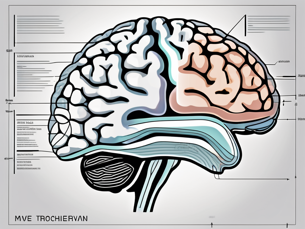
The trochlear nerve, also known as cranial nerve IV, is a vital component of the human nervous system. Situated within the brainstem, this nerve plays a crucial role in eye movement and coordination. Understanding the intricate workings of the trochlear nerve provides valuable insights into its anatomical structure, functions, associated disorders, and available treatment options.
Understanding the Trochlear Nerve
The trochlear nerve is a fascinating component of the human nervous system that plays a crucial role in eye movement and coordination. Let’s delve deeper into the anatomy and function of this remarkable cranial nerve.
Anatomy of the Trochlear Nerve
The trochlear nerve emerges from the dorsal aspect of the midbrain, specifically from the trochlear nucleus. This nucleus is located in the posterior part of the midbrain, near the cerebral aqueduct. It is here that the trochlear nerve begins its journey, carrying important signals that will ultimately control eye movement.
Unlike other cranial nerves, the trochlear nerve is unique in that it innervates a contralateral muscle, the superior oblique muscle. This means that the trochlear nerve controls the movement of the superior oblique muscle on the opposite side of the brain. The superior oblique muscle is responsible for a specific type of eye movement known as intorsion, which involves inward rotation of the eye.
After emerging from the trochlear nucleus, the trochlear nerve courses through the cavernous sinus, a cavity located within the skull. This sinus is an intricate network of veins and nerves that serves as a pathway for various structures, including the trochlear nerve. From there, the trochlear nerve enters the orbit through the superior orbital fissure, a narrow opening in the bony structure of the skull.
Once inside the orbit, the trochlear nerve reaches its destination – the superior oblique muscle. It influences the movement of this muscle, allowing for precise control over vertical and rotational eye movements. This intricate coordination between the trochlear nerve and the superior oblique muscle is essential for proper visual function.
Function of the Trochlear Nerve
The primary function of the trochlear nerve is to control the superior oblique muscle, which aids in one’s ability to move their eyes in a vertical and rotational manner. This enables the eyes to track moving objects, maintain proper alignment, and adjust the visual focus accordingly.
Working in harmony with the other cranial nerves involved in eye movement, the trochlear nerve plays a crucial role in overall visual coordination and perception. It ensures that our eyes can smoothly and accurately follow objects, allowing us to navigate our surroundings and interact with the world around us.
Without the trochlear nerve, our eye movements would be limited, hindering our ability to explore our environment and engage in daily activities. The intricate connections between the trochlear nerve, the superior oblique muscle, and the other cranial nerves involved in eye movement highlight the complexity and precision of the human visual system.
In conclusion, the trochlear nerve is a remarkable cranial nerve that emerges from the midbrain and controls the movement of the superior oblique muscle. Its role in eye movement coordination is vital for maintaining proper visual function and perception. Understanding the anatomy and function of the trochlear nerve allows us to appreciate the intricate mechanisms that enable us to see and interact with the world.
The Role of the Trochlear Nerve in Eye Movement
The trochlear nerve plays a crucial role in facilitating complex eye movements. It is responsible for coordinating the actions of the superior oblique muscle, which is one of the six extraocular muscles that control the movement of the eyes.
Trochlear Nerve and Superior Oblique Muscle
The trochlear nerve and the superior oblique muscle work synergistically to enable precise eye movements. When the trochlear nerve is stimulated, it triggers the contraction of the superior oblique muscle. This contraction leads to the downward rotation and inward rotation of the eyes, allowing for accurate depth perception and the ability to track objects moving in a superior and medial direction.
The superior oblique muscle plays a vital role in eye movement as it helps to counteract the actions of other eye muscles. It acts as a depressor, intorter, and abductor of the eye, working in opposition to the actions of the inferior oblique, superior rectus, and lateral rectus muscles, respectively.
Together, the trochlear nerve and the superior oblique muscle contribute to the intricate coordination required for smooth and accurate eye movements.
Impact on Vision and Eye Coordination
Imbalances or dysfunction of the trochlear nerve can have a significant impact on vision and eye coordination. Trochlear nerve palsy, for instance, can cause vertical or torsional diplopia (double vision), as well as difficulty in looking downwards or medially.
Vertical diplopia occurs when the eyes are unable to align properly, resulting in two images being perceived instead of one. This can lead to a distorted visual perception and difficulties in tasks that require depth perception, such as judging distances or catching objects. Torsional diplopia, on the other hand, refers to double vision with a rotational component, causing objects to appear tilted or slanted.
In addition to diplopia, trochlear nerve dysfunction can also affect eye movements, making it challenging to look downwards or medially. This limitation can hinder daily activities such as reading, driving, or participating in sports that require quick eye movements and changes in focus.
It is crucial to promptly identify and address any issues related to the trochlear nerve to minimize the impact on vision and overall quality of life. Treatment options may include medication, vision therapy, or in severe cases, surgical intervention to correct the underlying cause of the dysfunction.
In conclusion, the trochlear nerve and the superior oblique muscle work together to facilitate complex eye movements, allowing for precise depth perception and the ability to track objects. Dysfunction of the trochlear nerve can have a significant impact on vision and eye coordination, highlighting the importance of early detection and appropriate management.
Disorders Associated with the Trochlear Nerve
The trochlear nerve, also known as the fourth cranial nerve, plays a vital role in eye movement. It is responsible for controlling the superior oblique muscle, which helps rotate the eye downward and inward. When the trochlear nerve is affected by various disorders, it can lead to trochlear nerve palsy, causing significant visual disturbances and impairments.
Causes of Trochlear Nerve Palsy
Trochlear nerve palsy can occur due to several factors, with trauma and vascular events being the most common causes. Head injuries, such as those sustained in accidents or sports-related incidents, can damage the trochlear nerve and result in palsy. Additionally, ischemic strokes, caused by a blockage in the blood vessels supplying the brain, can also lead to trochlear nerve dysfunction.
Structural abnormalities, such as congenital malformations or acquired deformities, can affect the trochlear nerve’s normal functioning. Tumors, both benign and malignant, can also compress or invade the nerve, causing palsy. Furthermore, infections, such as meningitis or sinusitis, can lead to inflammation and damage to the trochlear nerve.
It is crucial to consult with a medical professional if you experience any symptoms of trochlear nerve palsy. They can provide an accurate diagnosis and determine the underlying cause, which is essential for developing an appropriate treatment plan. The treatment options may vary depending on the specific etiology, and early intervention can significantly improve the prognosis.
Symptoms and Diagnosis of Trochlear Nerve Disorders
Trochlear nerve disorders can present with a range of symptoms, primarily affecting eye movement and vision. One of the most common symptoms is double vision, also known as diplopia. This occurs because the affected eye is unable to move properly, leading to misalignment and overlapping of images.
Another characteristic symptom of trochlear nerve disorders is difficulty with downward gaze. Patients may experience a limited ability to look downward, making tasks such as reading or navigating stairs challenging. Tilting the head to one side or backward can alleviate the visual disturbances temporarily, as it compensates for the impaired eye movement.
Accurate diagnosis of trochlear nerve disorders typically involves a comprehensive evaluation by a healthcare professional. They will conduct a thorough medical history assessment, looking for any potential risk factors or previous incidents that may have contributed to the nerve dysfunction. A detailed ophthalmological examination will be performed to assess eye movement, alignment, and visual acuity.
In some cases, additional tests may be necessary to pinpoint the root cause and extent of the nerve dysfunction. These may include imaging studies, such as magnetic resonance imaging (MRI) or computed tomography (CT) scans, to visualize any structural abnormalities, tumors, or signs of inflammation. The results of these tests can provide valuable insights for the healthcare professional to make an accurate diagnosis and develop an appropriate treatment plan.
It is crucial not to self-diagnose or ignore any symptoms related to trochlear nerve disorders. Consulting with a qualified healthcare professional is essential for receiving appropriate medical guidance and ensuring the best possible outcomes. Early detection and intervention can significantly improve the prognosis and quality of life for individuals affected by trochlear nerve disorders.
Treatment Options for Trochlear Nerve Damage
Trochlear nerve damage can significantly impact eye movement and visual function. The treatment approach for this condition primarily depends on the underlying cause and severity of the damage. Non-surgical interventions are often the first line of treatment, focusing on managing modifiable risk factors and improving eye coordination. In some cases, surgical interventions may be necessary to repair or release the trochlear nerve, providing relief from associated symptoms.
Non-Surgical Treatments
Non-surgical treatments for trochlear nerve damage aim to address modifiable risk factors and improve visual function. One common approach is to optimize blood pressure control, as high blood pressure can contribute to nerve damage. By managing blood pressure through lifestyle modifications or medication, the strain on the visual system can be reduced, potentially alleviating symptoms.
Another non-surgical treatment option involves prescribing corrective lenses. This is particularly beneficial for individuals with refractive errors, such as nearsightedness or farsightedness, which can put additional strain on the eyes. By wearing the appropriate corrective lenses, the visual system can function more efficiently, minimizing the impact of trochlear nerve damage.
Physical therapy and eye exercises are also commonly recommended for individuals with trochlear nerve damage. These interventions aim to enhance eye coordination and improve visual function. Physical therapists or specialized eye therapists can guide patients through specific exercises and techniques designed to strengthen the eye muscles and improve eye movement control.
It is important to note that before initiating any non-surgical treatment regimen, it is crucial to consult with a healthcare professional. They will be able to assess the individual’s specific condition and provide personalized recommendations. Each case of trochlear nerve damage is unique, and individualized care is essential for optimal outcomes.
Surgical Interventions for Trochlear Nerve Repair
In cases where non-surgical treatments do not yield satisfactory results, surgical interventions may be necessary to repair or release the trochlear nerve. These surgical procedures aim to restore normal eye movement and alleviate associated symptoms.
The specific surgical approach for trochlear nerve repair depends on the underlying pathology and severity of the damage. One common surgical procedure is nerve decompression, which involves relieving pressure on the trochlear nerve to restore its function. This can be achieved by removing any compressive structures or releasing tight tissues that may be impeding the nerve’s ability to transmit signals properly.
Another surgical option is muscle surgery, which involves repositioning or adjusting the eye muscles to improve eye movement. This procedure can help correct any misalignment or weakness in the affected eye, allowing for better coordination and function.
In more severe cases, nerve grafting may be considered. This surgical technique involves taking a healthy nerve from another part of the body and using it to repair the damaged trochlear nerve. This can help restore nerve function and improve eye movement.
Consulting with a skilled ophthalmologist or neurosurgeon is crucial to determine the most appropriate surgical course of action for trochlear nerve repair. These specialists will evaluate the individual’s specific condition, consider the potential risks and benefits of surgery, and develop a personalized treatment plan.
In conclusion, the treatment options for trochlear nerve damage range from non-surgical interventions to surgical procedures. The choice of treatment depends on the underlying cause and severity of the damage. Non-surgical treatments focus on managing modifiable risk factors, prescribing corrective lenses, and engaging in physical therapy and eye exercises. Surgical interventions aim to repair or release the trochlear nerve, with procedures such as nerve decompression, muscle surgery, or nerve grafting. Consulting with a healthcare professional is crucial to determine the most appropriate treatment approach for each individual.
The Trochlear Nerve in the Wider Nervous System
Interactions with Other Cranial Nerves
The trochlear nerve does not function in isolation but interacts intricately with other cranial nerves within the central nervous system. Notably, it cooperates with the oculomotor nerve (cranial nerve III) and the abducens nerve (cranial nerve VI) to orchestrate harmonious eye movement. These cranial nerves collectively play a pivotal role in maintaining the intricate balance required for optimal visual coordination and depth perception.
The oculomotor nerve, also known as cranial nerve III, is responsible for controlling most of the eye’s movements. It innervates the majority of the extraocular muscles, including the superior rectus, inferior rectus, medial rectus, and inferior oblique muscles. These muscles work together with the trochlear nerve to ensure smooth and coordinated eye movements in various directions.
The abducens nerve, or cranial nerve VI, is primarily responsible for the lateral movement of the eye. It innervates the lateral rectus muscle, which is responsible for abduction, or moving the eye away from the midline. The trochlear nerve collaborates with the abducens nerve to ensure precise and controlled eye movements, allowing us to focus on objects at different distances and angles.
The Trochlear Nerve’s Role in Overall Nervous System Function
Beyond its specific involvement in eye movement, the trochlear nerve contributes to the overall function and connectivity of the nervous system. By integrating the complex signals from various cranial nerves, it aids in the seamless coordination of motor and sensory information related to vision. The trochlear nerve’s role extends beyond eye movement, highlighting its significance within the broader intricacies of the nervous system.
The trochlear nerve receives inputs from the visual cortex, which is responsible for processing visual information. These inputs help the trochlear nerve modulate eye movements based on the visual stimuli received. Additionally, the trochlear nerve interacts with other regions of the brain, such as the cerebellum, to ensure precise and coordinated eye movements.
Moreover, the trochlear nerve plays a crucial role in maintaining balance and spatial orientation. It receives inputs from the vestibular system, which is responsible for detecting changes in head position and movement. This integration of vestibular and visual information allows the trochlear nerve to adjust eye movements accordingly, ensuring that our vision remains stable even during head movements or changes in body position.
Furthermore, the trochlear nerve is involved in the regulation of pupil size. It receives inputs from the autonomic nervous system, specifically the parasympathetic and sympathetic divisions. These inputs help control the constriction and dilation of the pupil, allowing for optimal visual acuity in different lighting conditions.
In conclusion, the trochlear nerve plays a vital role in controlling eye movement and coordination. Dysfunction or damage to this nerve can cause significant visual disturbances, underscoring the importance of understanding its anatomy, functions, associated disorders, and treatment options. If you experience any symptoms related to the trochlear nerve or have concerns about your vision, it is crucial to consult with a qualified healthcare professional for appropriate diagnosis and guidance.
