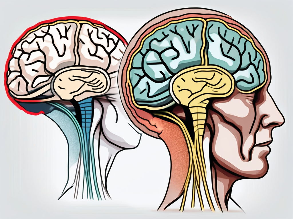
The topic of right trochlear nerve palsy can be quite complex and requires a thorough understanding of its anatomy, symptoms, diagnosis, and treatment options. In this article, we will delve into the various aspects of this condition, including the location of the lesion, the impact it has on the trochlear nerve, and the prognosis for patients affected by it.
Understanding Right Trochlear Nerve Palsy
Definition of Right Trochlear Nerve Palsy
Right trochlear nerve palsy is a neurological condition that affects the fourth cranial nerve, known as the trochlear nerve. The trochlear nerve is responsible for controlling the movement of the superior oblique muscle, which enables the eye to move downward and inward. When the trochlear nerve is damaged or impaired, it can lead to difficulties in controlling eye movement, specifically the ability to look downward and inward with the affected eye.
In order to fully comprehend the impact of right trochlear nerve palsy, it is important to delve into the intricate workings of the cranial nerves. The cranial nerves are a set of twelve nerves that emerge directly from the brain and are responsible for transmitting sensory and motor information to and from various parts of the head and neck. Each cranial nerve has a specific function, and any disruption or damage to these nerves can result in a range of neurological conditions.
The trochlear nerve, in particular, plays a crucial role in eye movement. Originating from the dorsal aspect of the midbrain, it is the only cranial nerve that emerges from the posterior aspect of the brainstem. Its unique pathway and function make it susceptible to injury or dysfunction.
Symptoms and Diagnosis
Symptoms of right trochlear nerve palsy may vary depending on the severity of the condition. Common signs include vertical diplopia (double vision), often worsened when looking downward or inward, as well as a tilting or torsional deviation of the affected eye. Patients may also experience headaches and eye strain due to the misalignment of the eyes.
Living with right trochlear nerve palsy can be challenging, as it affects not only visual perception but also daily activities such as reading, driving, and even walking. The constant struggle to align the eyes and overcome the double vision can lead to fatigue and frustration.
Diagnosing right trochlear nerve palsy involves a comprehensive evaluation by a medical professional, typically an ophthalmologist or neurologist. A thorough medical history, physical examination, and specialized tests are conducted to identify the specific cause and location of the lesion. These diagnostic tests may include a visual field examination, ocular motility testing, and imaging studies such as CT scans or MRI.
During the physical examination, the doctor may assess the patient’s eye movements, looking for any abnormalities or limitations in the affected eye. They may also perform a cover test, which involves covering one eye at a time to observe any misalignment or deviation. Additionally, the doctor may use prisms or other devices to determine the degree of double vision and assess the patient’s ability to fuse the images from both eyes.
Imaging studies, such as CT scans or MRI, are often used to identify the underlying cause of right trochlear nerve palsy. These tests can provide detailed images of the brain and cranial nerves, helping to pinpoint any structural abnormalities or lesions that may be affecting the trochlear nerve.
Once a diagnosis is made, the medical professional will work with the patient to develop an appropriate treatment plan. Treatment options for right trochlear nerve palsy may include the use of prism glasses to alleviate double vision, eye exercises to improve eye coordination, and in some cases, surgical intervention to correct any structural abnormalities or repair the damaged nerve.
It is important for individuals with right trochlear nerve palsy to seek medical attention and follow the recommended treatment plan to manage their symptoms effectively. With proper care and support, many individuals with this condition can regain functional vision and improve their quality of life.
Anatomy of the Trochlear Nerve
Location and Function of the Trochlear Nerve
The trochlear nerve, also known as the fourth cranial nerve, is a fascinating component of the human nervous system. It is the thinnest cranial nerve in the body and originates from the dorsal aspect of the midbrain. As it travels through the brain, it passes through the trochlear nucleus and trochlear fovea, which are essential structures involved in its function.
The name “trochlear” is derived from the Greek word “trochlea,” meaning “pulley.” This name is fitting because the trochlear nerve plays a crucial role in eye movements. Specifically, it innervates the superior oblique muscle, which is responsible for primarily depressing and intorting the eye. This muscle’s actions are particularly important when looking downward and inward, allowing us to navigate our visual environment with precision.
Disruption to the trochlear nerve can have significant consequences on eye movements. If the nerve is impaired, it can result in right trochlear nerve palsy, depending on the affected side. This condition can lead to difficulties in performing specific eye movements, affecting a person’s ability to focus and track objects accurately.
The Pathway of the Trochlear Nerve
The journey of the trochlear nerve is a remarkable one, spanning both intracranial and extracranial regions. After originating from the midbrain, the nerve decussates, meaning it crosses within the brainstem. This crossing allows for coordinated eye movements between the two sides of the body.
Once it has crossed, the trochlear nerve exits the brain through the posterior cranial fossa. From there, it embarks on a winding path around the midbrain, navigating through a complex venous structure known as the cavernous sinus. This sinus, located behind the eye, houses various blood vessels and nerves, including the trochlear nerve.
Finally, the trochlear nerve enters the orbit, the bony socket that houses the eye. It does so through a small opening called the superior orbital fissure. Once inside the orbit, the trochlear nerve innervates the superior oblique muscle, ensuring its proper function in eye movements.
Given its extensive intracranial and extracranial pathway, the trochlear nerve is susceptible to various potential sites of lesion or damage. Understanding the specific causes of these lesions is crucial in formulating an accurate prognosis and appropriate treatment plan for affected patients. It requires a comprehensive understanding of the intricate anatomy and physiology of the trochlear nerve.
In conclusion, the trochlear nerve is a remarkable cranial nerve that plays a vital role in eye movements. Its thin structure and intricate pathway make it susceptible to various potential sites of damage. By further exploring the anatomy and function of this nerve, researchers and healthcare professionals can continue to deepen their understanding of its complexities and develop innovative treatments for related conditions.
Lesion in Right Trochlear Nerve Palsy
Causes of Lesions in the Trochlear Nerve
Lesions within the trochlear nerve can occur due to various factors, including trauma, infections, tumors, vascular malformations, or autoimmune disorders. Traumatic causes, such as head injuries or fractures involving the skull base, can lead to damage or compression of the trochlear nerve. Infections, such as meningitis or sinusitis, can also affect the nerve, causing inflammation or direct damage.
Tumors, both benign and malignant, may impinge upon or infiltrate the trochlear nerve, resulting in its dysfunction. Vascular malformations, such as arteriovenous malformations or aneurysms, can disrupt the nerve’s blood supply, leading to ischemia and subsequent palsy. Additionally, certain autoimmune disorders, including multiple sclerosis, can cause demyelination of the nerve fibers, leading to impairment.
It is important to note that the trochlear nerve is the smallest cranial nerve and has the longest intracranial course. Due to its vulnerable position, it is susceptible to various pathologies that can result in lesions and subsequent palsy.
Impact of Lesions on the Trochlear Nerve
Lesions within the trochlear nerve can have a significant impact on a patient’s daily life, particularly in terms of visual function and ocular alignment. The loss of superior oblique muscle function in the affected eye results in difficulties looking downward and inward. This might affect tasks that require reading or viewing objects at close range.
In addition to the visual impairment, right trochlear nerve palsy can cause significant discomfort and frustration. Individuals may experience challenges with depth perception, leading to difficulties navigating stairs or judging distances accurately. The loss of binocular vision can also affect hand-eye coordination, making activities such as catching a ball or driving a car more challenging.
Furthermore, the trochlear nerve plays a crucial role in maintaining eye alignment. Lesions in the trochlear nerve can result in a misalignment of the eyes, known as strabismus. This misalignment can lead to double vision (diplopia) and may require interventions such as prism glasses or surgical correction to restore proper alignment.
It is important to note that the impact of the lesion can vary among patients, with some experiencing more severe symptoms than others. Factors such as the location and extent of the lesion, as well as the individual’s overall health and age, can influence the severity of the symptoms and the potential for recovery.
Treatment and Management of Right Trochlear Nerve Palsy
Medical Interventions for Trochlear Nerve Palsy
The treatment of right trochlear nerve palsy depends on the underlying cause, severity of symptoms, and individual patient characteristics. In some cases, conservative measures such as patching one eye or using prism glasses may be recommended to improve visual alignment and reduce double vision.
In other instances, medical interventions may be necessary. This can include medications to manage pain or inflammation, as well as specialized eye exercises or ocular muscle strengthening therapies. Surgical options, such as trochleoplasty or eye muscle surgery, may be considered for cases where the lesion is severe or causing significant functional limitations.
It is crucial to consult with a medical professional to determine the most appropriate treatment plan tailored to individual needs, as well as to understand the potential risks and benefits associated with each approach.
When it comes to managing trochlear nerve palsy, a multidisciplinary approach is often employed. This involves collaboration between ophthalmologists, neurologists, physical therapists, and occupational therapists. By working together, these healthcare professionals can ensure a comprehensive and holistic treatment plan that addresses the various aspects of the condition.
For patients with mild symptoms, non-invasive interventions may be sufficient to alleviate discomfort and improve visual function. However, for those with more severe cases, a combination of medical and surgical interventions may be necessary to achieve optimal outcomes.
Rehabilitation and Therapy Options
A comprehensive rehabilitation program can significantly benefit patients with right trochlear nerve palsy. Physical therapy and occupational therapy may be recommended to improve eye alignment, enhance eye movements, and address any associated muscular imbalances or proprioceptive deficits. These therapies can also help individuals adapt to the functional challenges caused by the palsy and improve overall quality of life.
In addition to traditional rehabilitation approaches, newer advancements in technology, such as virtual reality training and computer-based exercises, are showing promise in enhancing visual function and aiding in the recovery process. These innovative therapies can potentially optimize the visual outcomes for individuals affected by right trochlear nerve palsy.
Furthermore, psychological support and counseling may be beneficial for patients with trochlear nerve palsy. Coping with the physical and emotional challenges of the condition can be overwhelming, and having a support system in place can greatly improve the patient’s overall well-being.
It is important to note that the rehabilitation process for trochlear nerve palsy is highly individualized. Each patient’s treatment plan will be tailored to their specific needs, taking into account factors such as age, overall health, and the extent of nerve damage. Regular reassessment and adjustments to the treatment plan may be necessary to ensure ongoing progress and optimal outcomes.
Prognosis of Right Trochlear Nerve Palsy
Factors Influencing Recovery
The prognosis for right trochlear nerve palsy varies depending on the cause, severity of the lesion, and individual factors. In cases where the damage is mild and there is no underlying structural abnormality, recovery may occur spontaneously over time, with the nerve regenerating and function gradually returning.
However, if the lesion is more severe or due to a chronic condition like multiple sclerosis, full recovery may be less likely. The presence of comorbidities, age, and overall health can also influence the prognosis. A thorough evaluation by a medical professional is vital in determining the specific prognosis for each individual case.
When it comes to the recovery process, it is important to consider the body’s remarkable ability to heal itself. Nerves, including the trochlear nerve, have the potential to regenerate and repair. This process, known as neuroplasticity, allows the nervous system to adapt and compensate for damage. In the case of right trochlear nerve palsy, the body may gradually establish new connections and pathways to restore function.
Additionally, the body’s response to treatment can play a significant role in the prognosis. Physical therapy and exercises specifically targeting the affected eye muscles can aid in the recovery process. These exercises may involve eye movements, focusing exercises, and coordination drills. By consistently engaging in these therapeutic activities, patients can enhance their chances of regaining full or partial function.
Long-term Outlook for Patients with Right Trochlear Nerve Palsy
Living with right trochlear nerve palsy can present challenges, but with appropriate treatment and management, individuals can adapt and lead fulfilling lives. Some patients may continue to experience residual symptoms, such as mild diplopia or limitations in specific eye movements.
It is important to note that the long-term outlook for patients with right trochlear nerve palsy can vary greatly depending on the individual and the underlying cause of the condition. Some individuals may experience significant improvement over time, while others may have more persistent symptoms. Regular follow-up with a healthcare professional specializing in neurology or ophthalmology is essential to monitor any changes in symptoms, ensure optimal management, and adjust treatment strategies as needed.
Furthermore, it is crucial to address the emotional and psychological impact of living with right trochlear nerve palsy. Coping with a chronic condition can be challenging, and seeking emotional support is an important aspect of overall well-being. Connecting with support groups or online communities where individuals share similar experiences can provide valuable insights, advice, and a sense of belonging.
In conclusion, the location of the lesion in right trochlear nerve palsy can vary and is influenced by various factors. Understanding the anatomy, symptoms, and impact of the lesion is crucial in formulating an accurate diagnosis and effective treatment plan. Seeking professional medical advice is imperative to receive proper evaluation, guidance, and personalized care. By increasing awareness about this condition, we hope to improve the understanding and support available to patients living with right trochlear nerve palsy.
