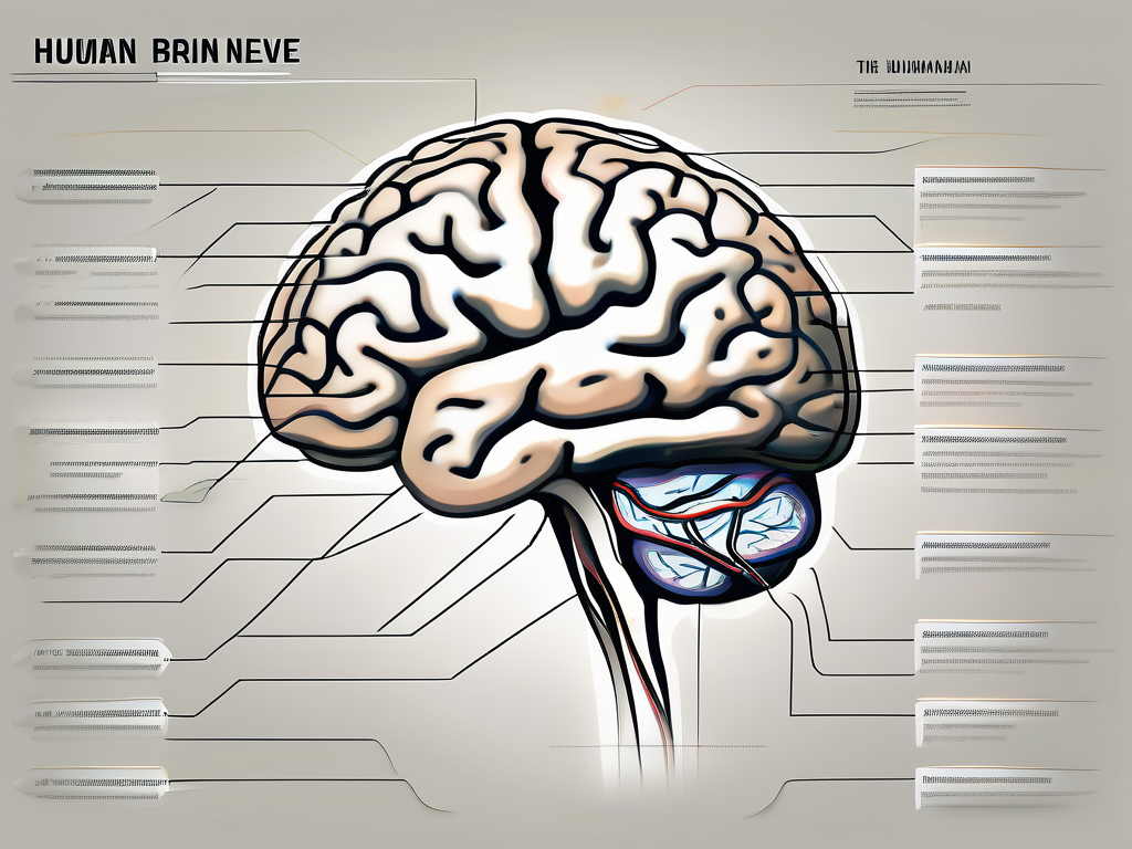
The trochlear nerve, also known as the fourth cranial nerve or CN IV, is a crucial component of the human nervous system. It plays an essential role in the complex mechanisms of vision and eye movement. Understanding the functions, anatomy, and disorders associated with the trochlear nerve can provide valuable insights into the intricate workings of the human body.
Understanding the Trochlear Nerve
The trochlear nerve is one of the twelve pairs of cranial nerves originating from the brainstem. It is labeled as the “fourth” cranial nerve due to its anatomical position. Unlike other cranial nerves, the trochlear nerve activates only one muscle, namely the superior oblique muscle, responsible for specific eye movements.
The trochlear nerve plays a crucial role in the complex system that allows us to see and perceive the world around us. By understanding its anatomy and functions, we can gain insights into the intricate mechanisms that govern our vision.
Anatomy of the Trochlear Nerve
The trochlear nerve emerges from the posterior aspect of the brainstem, specifically the midbrain. It courses along the surface and ultimately crosses the midline before entering the cavernous sinus.
From the cavernous sinus, the trochlear nerve enters the orbit through the superior orbital fissure, a narrow passageway behind the eye socket. It innervates the superior oblique muscle, which coordinates various eye movements, particularly downward and inward rotations.
The trochlear nerve’s path through the brainstem and orbit is a remarkable example of the intricate connections within our nervous system. It navigates through narrow passageways and interacts with other structures to ensure the precise control of eye movements.
Although the trochlear nerve is one of the smallest cranial nerves, its importance cannot be overstated. It plays a vital role in maintaining the delicate balance and coordination required for our eyes to function optimally.
Functions of the Trochlear Nerve
The primary function of the trochlear nerve is to enable coordinated eye movements. It controls the superior oblique muscle, which helps in the downward and inward rotation of the eye. This precise control allows for the alignment of both eyes, ensuring that images from the environment are properly focused on the retina for clear vision.
Without the trochlear nerve, our ability to move our eyes smoothly and accurately would be compromised. Simple tasks such as reading, tracking moving objects, and maintaining visual stability would become challenging.
In addition to its role in eye movements, the trochlear nerve also contributes to our sense of spatial awareness. By providing feedback to the brain about the position and orientation of the eyes, it helps us navigate our surroundings and perceive depth and distance accurately.
Disorders or injuries affecting the trochlear nerve can lead to a range of symptoms, including double vision, difficulty in looking downward, and an abnormal head tilt. These issues highlight the critical role that the trochlear nerve plays in maintaining the intricate balance of our visual system.
In conclusion, the trochlear nerve is a remarkable structure that enables precise control of eye movements. Its anatomy and functions are intricately connected to ensure optimal vision and spatial awareness. Understanding the trochlear nerve’s role in our visual system provides valuable insights into the complex mechanisms that govern our ability to see and perceive the world around us.
The Role of the Trochlear Nerve in Vision
Vision is an elaborate process that involves the integration of various components within the eye and the central nervous system. The trochlear nerve contributes significantly to this process through its influence on eye movements.
The trochlear nerve, also known as the fourth cranial nerve, is responsible for the innervation of the superior oblique muscle. This muscle plays a critical role in specific eye movements, ensuring that our visual system functions optimally.
Eye Movement and the Trochlear Nerve
The trochlear nerve works in conjunction with other cranial nerves to facilitate coordinated eye movements. It provides upward rotation of the eye when we look down or when our head tilts forward. This movement compensates for the natural tendency of the eyes to move in the opposite direction when viewing near objects.
Imagine you are reading a book. As your eyes move from line to line, the trochlear nerve ensures that your eyes rotate upwards slightly, allowing for a smooth transition between lines. Without the trochlear nerve’s contribution, reading would become a challenging task, as the eyes would constantly move in the opposite direction.
The synchronization of eye movements is vital for tasks such as reading, tracking moving objects, and depth perception. The trochlear nerve ensures that these movements occur seamlessly, allowing us to navigate our visual surroundings with ease.
The Trochlear Nerve and Superior Oblique Muscle
The superior oblique muscle, innervated by the trochlear nerve, plays a critical role in specific eye movements. This muscle is responsible for tendon pulley, which changes the direction of force by rotating the eye along its visual axis. It helps to tilt and depress the eye while also moving it inward.
Next time you gaze at a distant object, observe how your eyes move. The superior oblique muscle, under the control of the trochlear nerve, subtly adjusts the position of your eyes, allowing you to maintain a clear and focused view. This adjustment is especially important when viewing objects at different distances, as it helps to maintain visual acuity.
When the trochlear nerve is functioning optimally, the superior oblique muscle contributes to well-coordinated eye movements and allows for smooth visual tracking. Whether you are watching a bird soar through the sky or following the path of a bouncing ball, the trochlear nerve ensures that your eyes move in perfect harmony, providing you with a seamless visual experience.
Disorders Associated with the Trochlear Nerve
Like any other component in the human body, the trochlear nerve can be susceptible to various disorders that may impair its function. Damage or dysfunction of the trochlear nerve can result in distinct symptoms and require appropriate diagnosis and treatment.
The trochlear nerve, also known as the fourth cranial nerve, is responsible for the movement of the superior oblique muscle in the eye. This muscle helps control the rotation and downward movement of the eye. When the trochlear nerve is affected, it can lead to a range of symptoms and visual disturbances.
Symptoms of Trochlear Nerve Damage
When the trochlear nerve is damaged, individuals may experience double vision, difficulty focusing on near objects, and problems with eye movement coordination. This can significantly impact overall vision, making simple tasks such as reading or driving challenging.
Double vision, also known as diplopia, occurs when the eyes are unable to align properly, resulting in two images being seen instead of one. This can be particularly disorienting and can affect depth perception. Difficulty focusing on near objects, known as near vision impairment, can make tasks like reading or using a computer screen challenging.
Problems with eye movement coordination, known as ocular motility disorders, can cause the eyes to move in an uncoordinated manner. This can lead to difficulties in tracking moving objects or following a line of text while reading.
If you are experiencing any of these symptoms, it is essential to consult with a medical professional for a thorough evaluation and diagnosis. Early detection and treatment can help prevent further complications and improve overall visual function.
Diagnosis and Treatment of Trochlear Nerve Disorders
To diagnose trochlear nerve disorders, healthcare professionals may perform a comprehensive eye examination, including an assessment of eye movements and coordination. This may involve tracking the movement of the eyes, checking for any abnormalities in eye alignment, and evaluating the function of the superior oblique muscle.
In addition to a physical examination, imaging tests, such as CT scans or MRIs, can help identify any structural abnormalities or lesions affecting the trochlear nerve. These tests can provide detailed images of the brain and eye structures, allowing healthcare professionals to pinpoint the exact cause of the trochlear nerve dysfunction.
Treatment options for trochlear nerve disorders vary depending on the underlying cause and severity of symptoms. In some cases, medication may be prescribed to alleviate symptoms and reduce inflammation or swelling around the nerve. Vision therapy, which involves exercises and techniques to improve eye coordination and strengthen the affected muscles, may also be recommended.
In more severe cases or when conservative treatments fail to provide relief, surgical intervention may be necessary. Surgical procedures can involve repairing or repositioning the superior oblique muscle, removing any obstructions or lesions affecting the trochlear nerve, or addressing any underlying conditions contributing to the nerve dysfunction.
It is important to note that the success of treatment for trochlear nerve disorders depends on various factors, including the specific condition, the individual’s overall health, and the extent of nerve damage. Therefore, a personalized treatment plan should be developed in consultation with a healthcare professional specializing in ophthalmology or neurology.
The Trochlear Nerve in the Larger Nervous System
The trochlear nerve is an integral part of the larger nervous system, coordinating eye movements and contributing to visual perception. Its connection to other cranial nerves and the brain highlights the complexity of neural interactions within the human body.
The Trochlear Nerve’s Connection to the Brain
The trochlear nerve originates from the midbrain, specifically the trochlear nucleus. This nucleus contains the cell bodies of the trochlear nerve’s motor neurons, which play a crucial role in transmitting signals between the brain and the superior oblique muscle.
These motor neurons are responsible for the contraction of the superior oblique muscle, one of the six extraocular muscles that control eye movements. The trochlear nerve’s connection to this muscle allows for precise and coordinated movements, such as downward and inward eye rotations.
Furthermore, the trochlear nerve’s fibers decussate, or cross over, within the midbrain. This unique anatomical feature allows the trochlear nerve to control the contralateral superior oblique muscle. In other words, the trochlear nerve originating from the right midbrain controls the left superior oblique muscle, and vice versa.
Understanding the intricate connections between the trochlear nerve and other regions of the brain contributes to our overall knowledge of how the nervous system regulates eye movements and vision.
Interaction of the Trochlear Nerve with Other Cranial Nerves
The trochlear nerve works in conjunction with other cranial nerves, such as the oculomotor nerve, abducens nerve, and the optic nerve, to ensure synchronized eye movements. These cranial nerves collectively provide the necessary control and coordination for visual perception and a wide range of eye movements.
The oculomotor nerve, for instance, innervates the majority of the extraocular muscles, including the superior rectus, inferior rectus, and medial rectus muscles. The abducens nerve, on the other hand, controls the lateral rectus muscle responsible for outward eye movements.
The optic nerve, which carries visual information from the retina to the brain, works in tandem with the trochlear nerve to process and interpret visual stimuli. This collaboration between the trochlear nerve and other cranial nerves allows for smooth and accurate eye movements, enabling us to track objects, shift our gaze, and maintain visual stability.
By working together, these vital components of the nervous system allow us to perceive the world around us and navigate our visual environments effectively.
In conclusion, the trochlear nerve plays a crucial role in eye movements and visual perception. Understanding its anatomy, functions, and associated disorders can provide valuable insights into the complexities of the human nervous system. If you suspect any issues with your trochlear nerve or experience vision-related symptoms, it is essential to seek professional medical advice and guidance. A thorough evaluation by a healthcare professional can help determine the most appropriate course of treatment for your specific condition.
