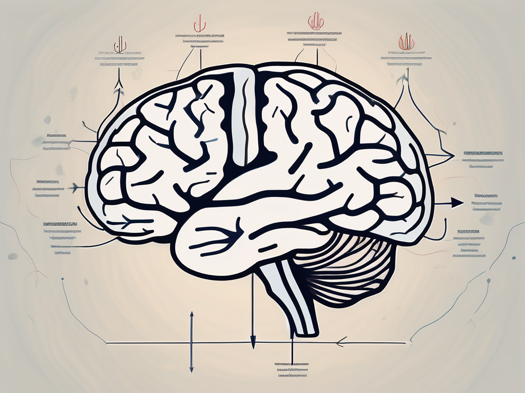
The trochlear nerve is one of the twelve cranial nerves responsible for the innervation of the muscles controlling eye movement. Dysfunction of the trochlear nerve can lead to various visual disturbances and physical discomfort. Understanding the causes, symptoms, and treatment options for trochlear nerve dysfunction can help individuals seek appropriate medical care and management.
Understanding the Trochlear Nerve
The trochlear nerve, also known as the fourth cranial nerve, is a critical component of the ocular motor system. This nerve originates from the brainstem, specifically the dorsal aspect of the tegmentum, and is the thinnest of all the cranial nerves. Its unique course extends around the brainstem and pierces the cavernous sinus before innervating the superior oblique muscle of the eye.
The trochlear nerve, with its delicate structure and intricate pathway, is a fascinating part of the human anatomy. Let’s delve deeper into its anatomy and function to gain a comprehensive understanding.
Anatomy of the Trochlear Nerve
The trochlear nerve emerges from the posterior aspect of the brainstem, opposite the midbrain. It traverses an intricate pathway dorsally and decussates within the superior medullary velum, a structure located within the fourth ventricle. Exiting the brainstem, the trochlear nerve enters the cavernous sinus and eventually reaches the superior oblique muscle of the eye, where it plays a crucial role in the coordination of vertical eye movements.
The journey of the trochlear nerve through the brainstem and cavernous sinus is a remarkable feat of nature’s design. Its precise route ensures that it reaches its target, the superior oblique muscle, with utmost accuracy.
Function of the Trochlear Nerve
The primary function of the trochlear nerve is to innervate the superior oblique muscle. This muscle enables the eye to move downward and inward, contributing to the depth perception and the ability to track objects in the visual field. Dysfunction of this nerve impairs the coordination of eye movements and can lead to a wide range of symptoms.
Imagine a scenario where the trochlear nerve is not functioning optimally. Tasks that require precise eye movements, such as reading or driving, become challenging. The ability to accurately judge distances and perceive depth is compromised, affecting daily activities that rely on visual perception.
Furthermore, the trochlear nerve’s role in coordinating vertical eye movements is crucial for maintaining balance and stability. When this nerve is impaired, individuals may experience difficulties in navigating stairs, walking on uneven surfaces, or even maintaining a steady gaze.
Understanding the trochlear nerve’s function allows us to appreciate the intricate mechanisms that enable our eyes to move with precision and accuracy. It highlights the importance of this small but mighty cranial nerve in our visual perception and overall well-being.
Common Symptoms of Trochlear Nerve Dysfunction
When the trochlear nerve is not functioning optimally, individuals may experience various visual disturbances and physical discomfort. It is essential to recognize these symptoms and seek medical evaluation for accurate diagnosis and treatment.
Visual Disturbances
One of the hallmark symptoms of trochlear nerve dysfunction is diplopia, commonly known as double vision. The affected individuals may report seeing two images instead of one, often overlapping or misaligned. The double vision is typically worse when looking downward or inward due to the impaired downward movement of the affected eye.
Double vision can significantly impact an individual’s daily life, making simple tasks such as reading, driving, or even walking challenging. The brain struggles to merge the two images into a single, coherent picture, leading to confusion and disorientation. This visual disturbance can cause frustration and anxiety, as individuals may find it difficult to navigate their surroundings confidently.
Moreover, the misalignment of the two images can cause eye strain and fatigue. The eyes work harder to focus and align the images, leading to discomfort and aching around the affected eye. This strain can further exacerbate the visual disturbances, making it even more challenging to perform visually demanding tasks.
Pain and Discomfort
In addition to visual disturbances, individuals may also experience pain and discomfort around the affected eye. This can manifest as headaches, eye strain, and a general sense of eye fatigue. The pain may worsen with eye movements or prolonged visual tasks, leading to further distress.
Headaches associated with trochlear nerve dysfunction can range from mild to severe, often presenting as a dull, throbbing pain around the temples or behind the eyes. The pain may be exacerbated by eye movements, such as looking up or down, and can persist for extended periods, affecting an individual’s overall well-being.
Eye strain is another common symptom experienced by individuals with trochlear nerve dysfunction. The eyes may feel tired, achy, or heavy, especially after prolonged visual tasks such as reading or using electronic devices. This discomfort can make it challenging to concentrate and maintain focus, impacting productivity and daily activities.
Furthermore, the general sense of eye fatigue can leave individuals feeling exhausted and drained. The constant effort required to compensate for the visual disturbances can take a toll on their energy levels, leading to decreased motivation and a decreased quality of life.
Causes of Trochlear Nerve Dysfunction
Trochlear nerve dysfunction can result from various underlying causes. Understanding these causes can help tailor appropriate treatment plans and preventive measures.
Let’s delve deeper into the different factors that can contribute to trochlear nerve dysfunction:
Trauma and Injury
Physical trauma, such as head injuries or fractures involving the orbit or skull base, can damage the trochlear nerve. The forceful impact can disrupt the delicate pathway of the nerve or cause swelling and inflammation in the surrounding tissues, leading to dysfunction.
For instance, a severe blow to the head during a car accident can result in a fracture of the skull base, affecting the trochlear nerve’s integrity. Similarly, a sports-related injury, like a direct hit to the eye, can cause trauma to the orbit and subsequently impact the nerve’s function.
It is important to note that the severity of the trauma and the extent of the damage can vary. In some cases, the trochlear nerve may only experience temporary dysfunction, while in others, the damage may be more severe and require long-term management.
Neurological Disorders
Certain neurological conditions, such as multiple sclerosis or brainstem lesions, can affect the trochlear nerve’s function. These disorders can disrupt the neural signals traveling along the nerve or affect the areas of the brainstem involved in its regulation.
Multiple sclerosis, for example, is an autoimmune disease that affects the central nervous system. In this condition, the body’s immune system mistakenly attacks the protective covering of nerve fibers, including those of the trochlear nerve. As a result, the nerve’s ability to transmit signals to the muscles responsible for eye movement becomes impaired.
Brainstem lesions, which can occur due to tumors or vascular malformations, can also disrupt the proper functioning of the trochlear nerve. These lesions can exert pressure on the nerve or interfere with the neural pathways, leading to dysfunction.
It is worth mentioning that neurological disorders affecting the trochlear nerve often present with a range of symptoms, including double vision, difficulty moving the affected eye, and eye misalignment.
Infections and Inflammations
Infections and inflammations, such as sinusitis or meningitis, can also contribute to trochlear nerve dysfunction. These inflammatory processes can impede the nerve’s proper functioning, leading to visual disturbances and discomfort.
Sinusitis, an inflammation of the sinuses, can cause swelling and pressure in the surrounding structures, including the trochlear nerve. The resulting compression can disrupt the nerve’s ability to transmit signals effectively, leading to eye movement abnormalities.
Meningitis, on the other hand, is an infection that causes inflammation of the protective membranes covering the brain and spinal cord. The inflammatory response can extend to the trochlear nerve, affecting its function. In severe cases, meningitis can lead to permanent damage to the nerve and subsequent visual impairments.
It is important to promptly diagnose and treat infections and inflammations that can affect the trochlear nerve to minimize the risk of long-term complications.
By understanding the various causes of trochlear nerve dysfunction, healthcare professionals can develop comprehensive treatment plans and preventive strategies tailored to each individual’s needs. Early intervention and appropriate management can significantly improve the prognosis and quality of life for those affected by this condition.
Diagnostic Techniques for Trochlear Nerve Dysfunction
Accurate diagnosis is crucial for determining the underlying cause of trochlear nerve dysfunction and guiding appropriate treatment strategies. Healthcare professionals employ several diagnostic techniques to evaluate the condition thoroughly.
Trochlear nerve dysfunction, also known as fourth cranial nerve palsy, can result in various symptoms such as double vision, difficulty in moving the eyes, and eye misalignment. To diagnose this condition, healthcare providers utilize a combination of clinical examination and imaging techniques.
Clinical Examination
A comprehensive clinical examination is the first step in assessing trochlear nerve dysfunction. Healthcare providers evaluate eye movements, visual acuity, and coordination. They carefully observe the patient’s ability to move their eyes in different directions, paying close attention to any abnormalities or limitations.
In addition to general eye movement assessment, healthcare providers may perform specialized tests to assess the degree of eye misalignment. One such test is the Maddox rod, which involves the use of a special lens that creates a visual illusion, helping to identify any deviations in eye alignment. Another test is the Hess screen, which uses a grid pattern to measure the extent of eye movement in different directions.
By conducting a thorough clinical examination, healthcare providers can gather valuable information about the patient’s symptoms and determine the severity of trochlear nerve dysfunction.
Imaging Techniques
To further investigate the underlying cause of trochlear nerve dysfunction, imaging techniques may be utilized. Magnetic resonance imaging (MRI) is commonly used to provide detailed images of the brain, cranial nerves, and surrounding structures. This non-invasive imaging modality helps identify any structural abnormalities, tumors, or lesions affecting the trochlear nerve.
During an MRI scan, the patient lies inside a large machine that uses a magnetic field and radio waves to create detailed cross-sectional images of the head and brain. These images can reveal any anatomical variations or pathological changes that may be contributing to the trochlear nerve dysfunction.
In some cases, healthcare providers may also use computed tomography (CT) scans to assess the bony structures of the skull and rule out any fractures or abnormalities that could be affecting the trochlear nerve.
By utilizing advanced imaging techniques, healthcare providers can obtain a clearer picture of the underlying cause of trochlear nerve dysfunction, enabling them to develop an appropriate treatment plan.
In conclusion, accurate diagnosis of trochlear nerve dysfunction involves a combination of clinical examination and imaging techniques. Through careful evaluation of eye movements, visual acuity, and coordination, healthcare providers can gather valuable information about the condition. Additionally, imaging techniques such as MRI and CT scans provide detailed images of the brain and surrounding structures, helping to identify any structural abnormalities or lesions affecting the trochlear nerve. This comprehensive approach to diagnosis ensures that appropriate treatment strategies can be implemented to address the underlying cause of trochlear nerve dysfunction.
Treatment Options for Trochlear Nerve Dysfunction
The management of trochlear nerve dysfunction depends on the underlying cause and severity of the condition. Treatment approaches aim to alleviate symptoms, improve visual function, and address the root cause of the dysfunction.
Trochlear nerve dysfunction, also known as fourth cranial nerve palsy, is a condition that affects the function of the trochlear nerve, which controls the movement of the superior oblique muscle in the eye. This can result in various symptoms, including double vision, difficulty looking downward, and eye misalignment.
When it comes to treating trochlear nerve dysfunction, healthcare professionals have several options at their disposal. These options can be broadly categorized into medications and therapies, as well as surgical interventions.
Medications and Therapies
Depending on the individual’s specific needs, healthcare professionals may recommend medications to manage pain, reduce inflammation, or control underlying neurological conditions. Nonsteroidal anti-inflammatory drugs (NSAIDs) may be prescribed to alleviate pain and reduce inflammation in the affected area. These medications can help provide temporary relief and improve the overall comfort of the patient.
In addition to medications, physical therapy and specialized eye exercises can also be prescribed to improve eye coordination and strengthen the affected muscles. Physical therapy sessions may include exercises that focus on eye movements, such as tracking objects or performing specific eye movements in different directions. These exercises aim to improve the coordination between the affected eye muscles and enhance overall visual function.
It is important to note that the effectiveness of these interventions can vary depending on the individual’s unique circumstances. Therefore, it is crucial to seek professional guidance from healthcare professionals who specialize in neurology or ophthalmology. These specialists can assess the severity of the trochlear nerve dysfunction and tailor the treatment plan to meet the specific needs of the patient.
Surgical Interventions
In some cases, surgical intervention may be necessary to address the underlying cause of trochlear nerve dysfunction. For instance, if trauma or structural abnormalities are responsible for the dysfunction, surgical repair or correction may be considered.
Surgical procedures for trochlear nerve dysfunction can vary depending on the specific cause and severity of the condition. One common surgical intervention is trochleoplasty, which involves reshaping the trochlea (a bony structure in the eye socket) to improve the alignment and movement of the superior oblique muscle.
It is crucial to consult with an experienced medical professional to evaluate the appropriateness of surgical intervention and discuss potential risks and benefits. The decision to undergo surgery should be made after a thorough assessment of the individual’s condition and consideration of alternative treatment options.
In conclusion, the treatment options for trochlear nerve dysfunction are diverse and depend on the underlying cause and severity of the condition. Medications and therapies, such as physical therapy and specialized eye exercises, can help alleviate symptoms and improve visual function. Surgical interventions may be necessary in certain cases to address structural abnormalities or trauma. It is important to consult with healthcare professionals who specialize in neurology or ophthalmology to determine the most appropriate treatment plan for each individual.
Prevention and Management of Trochlear Nerve Dysfunction
While not all instances of trochlear nerve dysfunction can be prevented, certain lifestyle modifications can potentially reduce the risk of injury or exacerbation. Additionally, regular check-ups and monitoring are essential for early detection and prompt intervention.
Lifestyle Modifications
Engaging in activities that promote eye safety, such as using protective eyewear during sports or occupational activities, can help minimize the risk of traumatic injuries to the eye and surrounding structures. Additionally, individuals should strive to maintain a healthy lifestyle, including regular exercise, a balanced diet, and adequate rest, to support overall eye health.
Regular Check-ups and Monitoring
Regular eye examinations allow healthcare professionals to monitor the function of the trochlear nerve and detect any abnormalities or changes in a timely manner. Routine check-ups are particularly crucial for individuals with underlying neurological conditions or a history of eye trauma. Any concerning symptoms should be promptly reported to healthcare providers for appropriate evaluation and management.
The Impact of Trochlear Nerve Dysfunction on Quality of Life
Trochlear nerve dysfunction can have a significant impact on an individual’s quality of life, both physically and emotionally. Understanding and addressing the potential consequences can help individuals seek appropriate support and take steps to manage their condition.
Physical Limitations
Visual disturbances, eye discomfort, and reduced eye movement coordination can restrict an individual’s ability to perform daily activities and affect their overall functionality. Physical limitations may include difficulties with reading, writing, driving, or engaging in sports or hobbies that require precise eye coordination. Seeking rehabilitation and appropriate management strategies can help individuals optimize their visual abilities and adapt to any physical limitations.
Psychological Effects
The impact of trochlear nerve dysfunction extends beyond physical limitations and can also affect an individual’s emotional well-being. Coping with visual disturbances, chronic pain, or a feeling of limitation in certain activities can lead to frustration, anxiety, or even depression. It is essential for individuals experiencing these psychological effects to seek appropriate support, such as counseling or support groups, in addition to medical management.
In conclusion, trochlear nerve dysfunction can give rise to various visual disturbances and physical discomfort. Familiarizing oneself with the causes, symptoms, and treatment options is essential for seeking timely medical evaluation and management. It is important to remember that this article provides general information and does not replace professional medical advice. If you suspect any trochlear nerve dysfunction or experience concerning symptoms, consult with a healthcare professional to receive personalized guidance and appropriate care.
