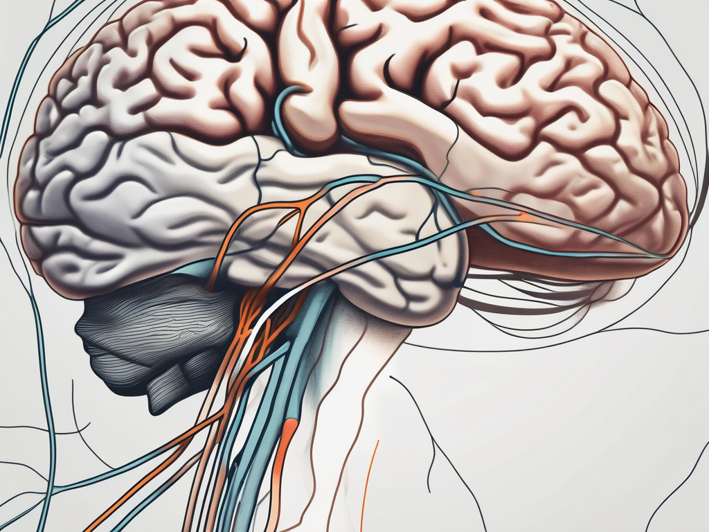
The trochlear nerve, also known as cranial nerve IV, is a vital component of the human nervous system. It plays a crucial role in facilitating efficient eye movement and control. Understanding the trochlear nerve’s anatomy, functions, and the disorders associated with it is essential for comprehending its significance in maintaining optimal vision.
Understanding the Trochlear Nerve
Anatomy of the Trochlear Nerve
The trochlear nerve is one of the twelve cranial nerves responsible for providing motor function to the eye muscles. It originates in the midbrain and is unique among the cranial nerves as it has the longest intracranial course.
As the trochlear nerve emerges from the dorsal aspect of the brainstem, it embarks on a fascinating journey through the intricate pathways of the nervous system. It navigates its way through a region called the posterior medullary velum, where it undergoes a remarkable process known as decussation. This means that the nerve fibers cross over to the opposite side of the brainstem, allowing for the precise coordination of eye movements.
After its adventurous crossing, the trochlear nerve enters the cavernous sinus, a complex network of veins situated at the base of the skull. This sinus serves as a protective conduit for the nerve, shielding it from potential damage and providing a stable environment for its passage.
Once the trochlear nerve completes its passage through the cavernous sinus, it continues its journey by traversing the superior orbital fissure, a bony opening in the skull. This opening acts as a gateway, allowing the nerve to reach its final destination – the superior oblique muscle of the eye.
The trochlear nerve’s arrival at the superior oblique muscle marks the culmination of its intricate pathway. It innervates the muscle with motor fibers, forming a vital connection that enables precise eye movement and coordination. The superior oblique muscle’s role in depression, or downward rotation, of the eye, as well as intorsion, or inward rotation, is made possible by the trochlear nerve’s motor function.
Functions of the Trochlear Nerve
The primary function of the trochlear nerve is to control the superior oblique muscle, which plays a pivotal role in specific types of eye movements. These movements include depression, or downward rotation, of the eye, as well as intorsion, or inward rotation.
By innervating the superior oblique muscle, the trochlear nerve allows for enhanced depth perception. This ability to accurately perceive depth is crucial for our everyday activities, such as judging distances and navigating our surroundings. Without the trochlear nerve’s contribution, our visual experience would lack the precision required for these tasks.
In addition to its role in depth perception, the trochlear nerve also aids in stabilizing visual images during head movement. This is achieved through the coordination of counter-rotational actions between both eyes. By working in harmony, the trochlear nerve ensures that our vision remains clear and focused, preventing excessive blurring or double vision.
Overall, the trochlear nerve’s intricate anatomy and essential functions make it a fascinating component of the cranial nerves. Its ability to control the superior oblique muscle and contribute to depth perception and visual stability highlights its importance in our daily visual experiences.
The Role of the Trochlear Nerve in Vision
The trochlear nerve plays a crucial role in vision by controlling the movements of the eye. It is responsible for coordinating the actions of several muscles, including the superior oblique muscle, which allows for specific eye movements such as rotation and elevation. These movements are essential for tracking moving objects or scanning our surroundings.
When we engage in activities like reading, driving, or playing sports, our eyes rely heavily on the coordinated actions of the trochlear nerve and the superior oblique muscle. Any changes or abnormalities in these eye movements can result in visual disturbances and difficulties in our daily activities. Therefore, it is important to carefully evaluate and address any issues related to the trochlear nerve’s function with the help of a medical professional.
Eye Movement and the Trochlear Nerve
Eye movements are fascinating processes that involve the intricate coordination of various muscles and nerves. The trochlear nerve, in particular, plays a significant role in these movements. It works in harmony with other eye muscles to ensure smooth and precise eye movements.
Imagine you are reading a book. As your eyes move across the page, the trochlear nerve sends signals to the superior oblique muscle, instructing it to rotate and elevate the eye. This allows you to track the words on the page effortlessly. Similarly, when you are driving, the trochlear nerve helps you scan the road ahead, ensuring that your eyes move smoothly from one point to another.
Without the trochlear nerve’s proper functioning, these eye movements can be disrupted. This can lead to difficulties in focusing, tracking objects, or maintaining proper eye alignment. For example, if the trochlear nerve is impaired, you may experience challenges in following a moving object, such as a ball during a sports game, or you may find it hard to read a line of text without losing your place.
The Trochlear Nerve and Superior Oblique Muscle
The trochlear nerve and the superior oblique muscle have a close relationship when it comes to eye alignment and positioning. The superior oblique muscle is responsible for rotating the eye downward and away from the midline, as well as intorting the eye (rotating it inward).
When the trochlear nerve functions properly, it sends signals to the superior oblique muscle, allowing it to contract and perform its intended actions. However, disorders involving the trochlear nerve can impact the superior oblique muscle’s ability to function effectively.
One such disorder is trochlear nerve palsy, which occurs when the trochlear nerve is weakened or paralyzed. In this condition, the superior oblique muscle may not receive the necessary signals to contract, resulting in visual impairments. Individuals with trochlear nerve palsy may experience double vision, difficulty looking downward, or a tilt in the affected eye.
It is important to note that trochlear nerve palsy can have various causes, including trauma, infections, or underlying medical conditions. Proper diagnosis and treatment are essential to address the underlying cause and manage the associated visual impairments effectively.
Disorders Associated with the Trochlear Nerve
The trochlear nerve, also known as the fourth cranial nerve, plays a vital role in eye movement. It innervates the superior oblique muscle, which is responsible for moving the eye downward and inward. Disorders associated with the trochlear nerve can lead to various symptoms and complications.
Causes of Trochlear Nerve Palsy
Trochlear nerve palsy can occur due to several factors, including trauma, infections, tumors, or vascular conditions affecting the nerve’s blood supply. Head injuries, such as skull fractures or concussions, can damage the trochlear nerve and impair its ability to transmit electrical impulses efficiently.
Similarly, tumors or infections in the brain or skull base can exert pressure on the nerve, disrupting its function. In some cases, trochlear nerve palsy may be congenital, meaning it is present at birth due to abnormalities in the development of the nerve.
If you experience any symptoms of trochlear nerve palsy, such as eye misalignment or visual disturbances, it is crucial to consult with a healthcare professional. They can conduct a thorough examination and diagnostic tests to pinpoint the underlying cause and develop an appropriate treatment plan.
Symptoms and Diagnosis of Trochlear Nerve Disorders
The symptoms of trochlear nerve disorders can vary depending on the specific condition and the individual affected. Common symptoms include double vision, eye misalignment, difficulty moving the eyes in a downward direction, and headaches.
A comprehensive eye examination, along with a detailed medical history review, is essential for diagnosing trochlear nerve disorders. During the eye examination, the healthcare professional will assess eye movements, alignment, and visual acuity. They may also perform additional tests, such as the Hess screen test, to evaluate the function of the extraocular muscles.
In some cases, additional tests such as imaging scans or electrophysiological studies may be required to confirm the diagnosis. Magnetic resonance imaging (MRI) or computed tomography (CT) scans can help identify any structural abnormalities or tumors affecting the trochlear nerve. Electrophysiological studies, such as electroretinography (ERG) or electrooculography (EOG), can provide valuable information about the electrical activity of the eye and its associated nerves.
Therefore, seeking early medical attention is crucial to ensure prompt diagnosis and appropriate intervention. Early detection and treatment can help prevent further complications and improve the overall prognosis for individuals with trochlear nerve disorders.
Treatment and Recovery for Trochlear Nerve Damage
Medical Interventions for Trochlear Nerve Disorders
The treatment options for trochlear nerve disorders depend on the cause, severity, and accompanying symptoms. Conservative management approaches, including visual aids and physical therapy, may be sufficient for individuals with mild trochlear nerve damage. Visual aids such as prism lenses can help correct double vision, while physical therapy can improve eye muscle strength and coordination.
When it comes to visual aids, prism lenses are a popular choice among healthcare professionals. These lenses work by bending light in a way that compensates for the misalignment of the eyes caused by trochlear nerve damage. By redirecting the light, prism lenses can help individuals with trochlear nerve disorders achieve clearer and more focused vision.
In addition to visual aids, physical therapy plays a crucial role in the treatment of trochlear nerve damage. Through a series of exercises and techniques, physical therapists can help patients improve their eye muscle strength and coordination. This can lead to better control over eye movements and a reduction in double vision.
In more severe cases, surgical interventions may be necessary to restore or improve the function of the trochlear nerve or the associated eye muscles. Surgical techniques can involve repositioning or strengthening the affected muscles, as well as decompressing or repairing the nerve itself.
One surgical approach commonly used for trochlear nerve disorders is muscle repositioning. This procedure involves moving the affected eye muscles to a new position, which can help alleviate the strain and improve their function. Another surgical technique is muscle strengthening, where the weakened eye muscles are strengthened through various methods, such as suturing or grafting.
For cases where the trochlear nerve itself is damaged, nerve decompression or repair may be necessary. Nerve decompression involves relieving any pressure or compression on the nerve, allowing it to function properly. Nerve repair, on the other hand, focuses on repairing any damage or injury to the nerve, which can help restore its function.
Prognosis and Recovery from Trochlear Nerve Damage
The prognosis for individuals with trochlear nerve damage depends on various factors, including the cause and severity of the injury, as well as the individual’s overall health. Prompt diagnosis and appropriate treatment interventions can significantly enhance the chances of recovery and restoration of visual function.
Recovery from trochlear nerve damage can be a gradual process. It may take time for the nerves and muscles to heal and regain their function fully. During this recovery period, individuals may continue to experience some symptoms, such as double vision or difficulty with eye movements. However, with proper treatment and rehabilitation, these symptoms can improve over time.
After treatment, diligent follow-up care and regular monitoring are crucial to ensure long-term visual health. It is important to maintain open communication with healthcare professionals, adhere to their recommendations, and promptly seek medical attention if any new symptoms or concerns arise.
Regular check-ups and eye examinations can help detect any potential complications or changes in visual function. These appointments also provide an opportunity for healthcare professionals to assess the effectiveness of the treatment and make any necessary adjustments to the management plan.
Additionally, individuals with trochlear nerve damage may benefit from ongoing support and rehabilitation services. This can include continued physical therapy sessions to further improve eye muscle strength and coordination, as well as counseling or support groups to address any emotional or psychological challenges that may arise during the recovery process.
Overall, with the right treatment approach and proper care, individuals with trochlear nerve damage can experience significant improvements in their visual function and quality of life.
In Conclusion
The trochlear nerve plays a crucial role in eye movement and coordination, contributing to our ability to perceive the world around us accurately. Understanding the anatomy, functions, and disorders associated with this vital cranial nerve is paramount in appreciating its significance in maintaining optimal vision.
If you experience any visual disturbances or suspect trochlear nerve-related issues, it is essential to consult with a healthcare professional. They possess the expertise and knowledge necessary to provide accurate diagnosis and guidance, leading to appropriate treatment and potentially restoring normal visual function.
