The trochlear nerve plays a crucial role in eye movement and coordination. However, when this nerve is damaged, various complications can arise. Understanding the anatomy and function of the trochlear nerve is essential in comprehending the impact of its damage. Additionally, identifying the causes, symptoms, and available treatment options are integral in managing trochlear nerve damage. This article explores these aspects, providing valuable insights into what happens when the trochlear nerve is damaged.
Understanding the Trochlear Nerve
Anatomy of the Trochlear Nerve
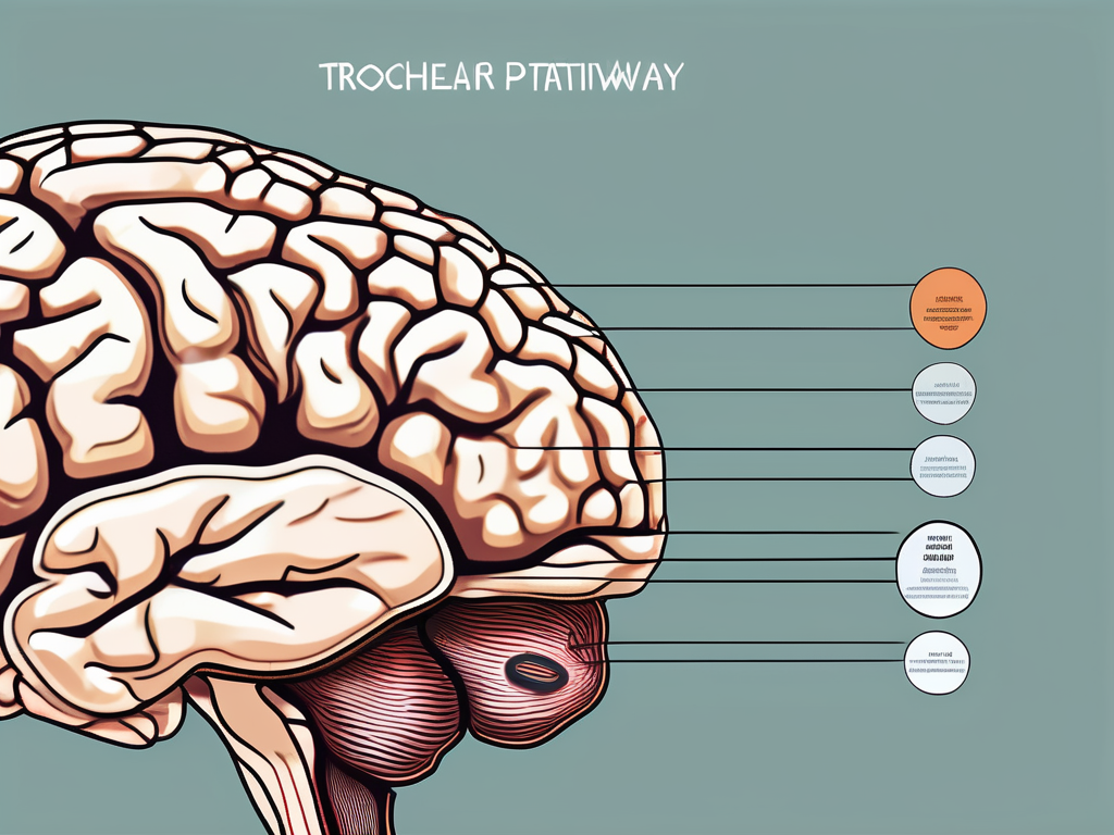
The trochlear nerve, also known as cranial nerve IV, is a motor nerve responsible for innervating the superior oblique muscle of the eye. It emerges from the brainstem, specifically the midbrain, and travels along a complex pathway before reaching its target. The unique trajectory of the trochlear nerve makes it susceptible to injury compared to other cranial nerves.
As the trochlear nerve emerges from the midbrain, it decussates, or crosses over, to the opposite side of the brainstem. This decussation occurs at the level of the superior colliculus, a structure involved in visual processing. From there, the trochlear nerve travels along the lateral aspect of the brainstem, passing through the cavernous sinus, a venous structure located on each side of the sella turcica, a bony depression in the sphenoid bone.
Continuing its journey, the trochlear nerve enters the orbit through the superior orbital fissure, a narrow opening located in the sphenoid bone. Once inside the orbit, it courses along the superior aspect of the medial wall, where it finally reaches its target, the superior oblique muscle.
Function of the Trochlear Nerve
The primary function of the trochlear nerve is to control the superior oblique muscle, which facilitates downward and inward eye movement. This muscle allows for proper eye alignment and coordinated gaze. When the trochlear nerve is damaged, these essential eye movements can be impaired, causing significant visual disturbances and discomfort for individuals.
The superior oblique muscle plays a crucial role in eye movements. It acts to depress, abduct, and internally rotate the eye. This complex set of actions allows for precise control of the visual field and helps maintain proper binocular vision. Without the trochlear nerve’s innervation, the superior oblique muscle would be unable to carry out its functions effectively, leading to difficulties in tasks that require accurate eye movements, such as reading, driving, and playing sports.
Injuries to the trochlear nerve can occur due to various reasons, including trauma, tumors, and vascular disorders. Depending on the severity of the damage, individuals may experience symptoms such as double vision, eye misalignment, and difficulty looking downward or inward. Treatment options for trochlear nerve injuries depend on the underlying cause and may include medication, surgery, or vision therapy to help improve eye coordination and function.
Causes of Trochlear Nerve Damage
Trauma and Injuries
Trochlear nerve damage can occur due to various traumatic incidents such as head trauma, fractures involving the orbit, or direct injury to the eye. The impact from a forceful blow can disrupt the nerve structure or cause it to stretch, leading to dysfunction.
Head trauma, especially when it involves a strong impact to the face or skull, can have devastating effects on the delicate trochlear nerve. The force of the blow can cause the nerve to be compressed or even severed, resulting in impaired eye movement and coordination.
Fractures involving the orbit, which is the bony socket that holds the eye, can also lead to trochlear nerve damage. When the bones around the eye are fractured, they can exert pressure on the nerve, causing it to become compressed or injured.
Direct injury to the eye, such as a penetrating injury or a foreign object striking the eye, can directly damage the trochlear nerve. The nerve fibers may be torn or stretched, disrupting the normal transmission of signals between the eye muscles and the brain.
Neurological Disorders
Certain neurological conditions, such as multiple sclerosis or stroke, can result in trochlear nerve damage. These disorders often affect the nervous system, compromising the transmission of signals along the nerve pathways. The trochlear nerve is not exempt from these effects.
Multiple sclerosis, a chronic autoimmune disease, can cause inflammation and damage to the protective covering of nerve fibers, known as myelin. When the myelin is damaged, the trochlear nerve may be affected, leading to problems with eye movement and coordination.
Stroke, which occurs when the blood supply to the brain is interrupted, can also have implications for the trochlear nerve. The lack of blood flow can result in damage to the nerve cells, impairing their ability to transmit signals effectively.
Other neurological disorders, such as tumors or infections affecting the brain or nervous system, can indirectly impact the trochlear nerve. The presence of these conditions can disrupt the normal functioning of the nerve pathways, leading to trochlear nerve damage.
Surgical Complications
In some cases, surgical procedures involving the eye may inadvertently damage the trochlear nerve. This can happen during interventions such as eye muscle surgery, orbital surgeries, or procedures aimed at addressing eye misalignment. Surgeons take great care to prevent damage to the trochlear nerve, but unforeseen circumstances can occur.
Eye muscle surgery, which is performed to correct strabismus (misalignment of the eyes), involves manipulating the eye muscles to improve their coordination. However, during the procedure, there is a risk of unintentionally damaging the trochlear nerve, which is responsible for controlling the superior oblique muscle.
Orbital surgeries, which involve interventions within the bony socket that holds the eye, can also pose a risk to the trochlear nerve. The close proximity of the nerve to the surgical site increases the chances of accidental injury during these procedures.
While surgeons take every precaution to avoid damaging the trochlear nerve, unforeseen circumstances can sometimes lead to surgical complications. Factors such as anatomical variations or unexpected bleeding can increase the risk of trochlear nerve damage during eye surgeries.
Symptoms of Trochlear Nerve Damage
Trochlear nerve damage can have a significant impact on an individual’s vision and overall quality of life. In addition to the common symptoms of double vision, blurred vision, and difficulty focusing, there are other aspects of this condition that deserve attention.
Vision Problems
Individuals with trochlear nerve damage often experience visual disturbances that go beyond the typical symptoms. In some cases, objects may not only appear double, but they may also seem tilted or shifted due to the misalignment of the affected eye. This misalignment can create a disorienting effect, making it difficult for individuals to accurately perceive their surroundings.
Furthermore, the impact of these visual difficulties can extend beyond the physical realm. Tasks that were once effortless, such as reading or driving, can become challenging and even dangerous. Everyday activities like walking down stairs may require extra caution and concentration, as the eye movement abnormalities associated with trochlear nerve damage can make it difficult to look downwards or inwards.
Eye Movement Difficulties
Eye movement abnormalities are one of the hallmark symptoms of trochlear nerve damage. The affected eye may appear misaligned or deviated, which can further exacerbate the functional impairment. The inability to move the eye in certain directions can significantly limit an individual’s range of vision and hinder their ability to perform daily tasks.
Imagine the frustration of not being able to comfortably read a book or watch a movie without experiencing discomfort or strain. These eye movement difficulties can have a profound impact on an individual’s independence and overall quality of life.
Pain and Discomfort
In addition to the visual and eye movement difficulties, trochlear nerve damage can also cause pain and discomfort around the affected eye. Headaches, eye strain, and a feeling of pressure behind the eye are common symptoms experienced by individuals with this condition.
These symptoms can be debilitating, affecting an individual’s overall well-being and productivity. The constant pain and discomfort can make it challenging to concentrate on tasks and may even lead to a decrease in social interactions and participation in activities that were once enjoyed.
It is important for individuals experiencing these symptoms to seek medical attention and receive a proper diagnosis. Understanding the full extent of trochlear nerve damage and its impact on vision and daily life can help healthcare professionals develop effective treatment plans and support strategies.
Diagnosing Trochlear Nerve Damage
Medical History and Physical Examination
When diagnosing trochlear nerve damage, healthcare professionals often begin by taking a detailed medical history and conducting a thorough physical examination. Patient-reported symptoms, past medical conditions, and recent injuries are all considered in the diagnostic process. A comprehensive eye examination is also essential in evaluating eye alignment, visual acuity, and eye movement.
During the medical history interview, the healthcare professional will ask the patient about any specific symptoms they are experiencing, such as double vision, difficulty moving the eyes, or pain around the eye area. They will also inquire about any previous medical conditions or injuries that may be relevant to the trochlear nerve damage.
The physical examination involves a careful assessment of the patient’s eye movements, focusing ability, and eye alignment. The healthcare professional may use specialized tools, such as a penlight or an ophthalmoscope, to examine the structures of the eye in more detail. They will observe the patient’s ability to follow objects with their eyes, check for any abnormalities in eye alignment, and assess visual acuity.
Imaging Tests
In some cases, imaging tests, such as magnetic resonance imaging (MRI) or computed tomography (CT) scans, may be ordered to visualize the structures surrounding the trochlear nerve. These imaging modalities can provide valuable information about potential structural abnormalities or lesions affecting the nerve.
An MRI uses powerful magnets and radio waves to create detailed images of the brain and surrounding structures. It can help identify any tumors, vascular malformations, or other abnormalities that may be compressing or damaging the trochlear nerve. A CT scan, on the other hand, uses X-rays to produce cross-sectional images of the head and can also provide valuable information about the bony structures and potential injuries.
Before undergoing an imaging test, the patient may be asked to remove any metal objects, such as jewelry or hairpins, as they can interfere with the magnetic field of the MRI machine. The patient will then be positioned on a table that slides into the scanner, which may cause some loud noises during the procedure. It is important for the patient to remain still during the imaging process to ensure clear and accurate images.
Neurological Evaluation
A neurological evaluation is often conducted to assess the overall function of the nervous system and rule out potential underlying neurological disorders. This evaluation may involve a series of tests to assess reflexes, coordination, and sensory perception.
During the neurological evaluation, the healthcare professional will assess the patient’s reflexes by tapping specific areas of the body with a reflex hammer. They will also evaluate the patient’s coordination by asking them to perform specific movements, such as touching their nose with their finger or walking in a straight line. Additionally, sensory perception tests may be conducted to assess the patient’s ability to feel different sensations, such as light touch or pinpricks.
Based on the findings from the medical history, physical examination, imaging tests, and neurological evaluation, healthcare professionals can make a more accurate diagnosis of trochlear nerve damage. This comprehensive approach ensures that all relevant factors are considered and helps guide appropriate treatment options for the patient.
Treatment Options for Trochlear Nerve Damage
Trochlear nerve damage can have a significant impact on a person’s quality of life, affecting their ability to move their eyes and causing discomfort. Fortunately, there are various treatment options available to manage the symptoms and improve eye movement coordination.
Medication and Drug Therapy
Depending on the underlying cause and severity of trochlear nerve damage, medication may be prescribed to manage associated symptoms. Pain relievers, such as nonsteroidal anti-inflammatory drugs (NSAIDs), can help alleviate discomfort. These medications work by reducing inflammation and relieving pain, allowing individuals to experience relief from the symptoms caused by trochlear nerve damage.
In addition to pain relief, lubricating eye drops or ointments may be recommended to relieve dryness or irritation. Trochlear nerve damage can lead to dry eyes, which can cause discomfort and affect vision. Lubricating eye drops or ointments help to keep the eyes moist, reducing dryness and irritation.
Physical Therapy and Rehabilitation
In cases where trochlear nerve damage affects eye movement coordination, physical therapy and rehabilitation may be beneficial. Eye exercises and specialized techniques can assist in improving eye muscle strength, eye tracking abilities, and visual coordination.
Physical therapists and rehabilitation specialists can design personalized exercise programs to target specific eye muscles and improve their function. These exercises may involve tracking objects with the eyes, focusing on different distances, and performing eye movements in various directions. Through consistent practice and guidance from professionals, individuals with trochlear nerve damage can regain control over their eye movements and improve their overall visual coordination.
Surgical Interventions
In severe cases of trochlear nerve damage or when non-surgical interventions prove ineffective, surgical procedures may be considered. Surgical options vary depending on the specific condition and may include muscle repositioning, nerve grafts, or other techniques aimed at restoring normal eye movement and function.
Before considering surgery, it is vital for individuals to consult with a qualified ophthalmologist or neurosurgeon. These specialists can assess the severity of the trochlear nerve damage and discuss the potential risks and benefits of surgical interventions. They will carefully evaluate the individual’s overall health and medical history to determine the most appropriate surgical approach.
Surgical interventions for trochlear nerve damage require expertise and precision, as they involve delicate structures in the eye and surrounding areas. The success of the surgery depends on the skill of the surgeon and the individual’s ability to recover and rehabilitate after the procedure.
In conclusion, treatment options for trochlear nerve damage include medication and drug therapy, physical therapy and rehabilitation, as well as surgical interventions. The choice of treatment depends on the underlying cause and severity of the condition. It is essential for individuals to work closely with healthcare professionals to determine the most suitable treatment plan and achieve the best possible outcomes.
Living with Trochlear Nerve Damage
Coping Mechanisms and Strategies
Living with trochlear nerve damage can be challenging, but there are coping mechanisms and strategies that can improve day-to-day life. Simple adjustments, such as using proper lighting, practicing eye hygiene, and implementing ergonomic strategies, can help alleviate symptoms and enhance visual comfort. Additionally, support groups and counseling services can provide emotional support and guidance to individuals navigating the challenges of trochlear nerve damage.
Support and Resources
It is imperative for individuals with trochlear nerve damage to seek support and resources that can assist in their overall well-being. Consulting with healthcare professionals, such as ophthalmologists or neurologists, can provide valuable guidance tailored to individual needs. Patient advocacy organizations and online forums can also serve as valuable sources of information and support.
Prognosis and Future Research
The prognosis for trochlear nerve damage varies depending on the underlying cause, severity, and timely intervention. With appropriate treatment, many individuals can experience improvements in their symptoms and quality of life. Ongoing research in the field of neurology and ophthalmology continues to shed light on innovative treatment modalities and management strategies for trochlear nerve damage, offering hope for future advancements in care.
In conclusion, trochlear nerve damage can lead to significant visual impairment and discomfort. Understanding the causes, symptoms, diagnosis, and available treatments is essential in managing this condition effectively. Individuals experiencing trochlear nerve damage should consult with healthcare professionals for proper evaluation, diagnosis, and personalized treatment plans. Early intervention and appropriate care can make a substantial difference in managing the impact of trochlear nerve damage and improving overall well-being.
