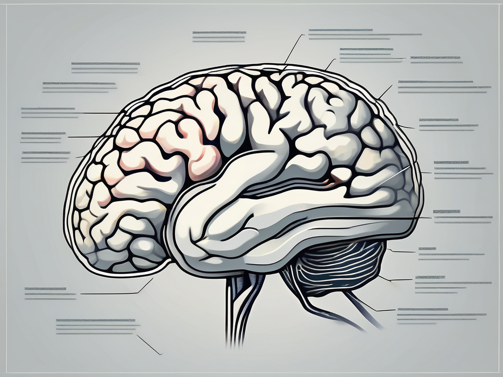
The trochlear nerve, also known as the fourth cranial nerve, plays a crucial role in the complex system of the human nervous system. Understanding its function is essential to comprehend its significance in maintaining various physiological processes and in maintaining overall health.
Understanding the Trochlear Nerve
In order to gain a comprehensive understanding of the trochlear nerve, it is important to delve into the intricacies of its anatomy and its role within the broader nervous system.
The trochlear nerve, also known as the fourth cranial nerve, derives its name from the Latin word “trochlea,” meaning a pulley. This name is fitting as it is responsible for the movement of the eye, acting like a pulley system to control the superior oblique muscle.
The anatomy of the trochlear nerve is fascinating. It primarily originates from the posterior aspect of the brainstem, specifically the midbrain. Unlike other cranial nerves, the trochlear nerve only contains motor fibers, making it unique. These motor fibers are responsible for carrying signals from the brain to the superior oblique muscle, allowing for precise eye movement.
Passing through the superior orbital fissure, a small opening in the skull, the trochlear nerve extends to the ocular orbit, where it innervates the superior oblique muscle. Its specific path and unique properties distinguish it from other cranial nerves, highlighting its specialized role in eye movement.
Anatomy of the Trochlear Nerve
The trochlear nerve originates from the trochlear nucleus, a small group of neurons located in the midbrain. From there, it travels dorsally, crossing the midline and decussating, meaning that the nerve fibers from one side of the brainstem cross over to the opposite side. This decussation is what allows the trochlear nerve to control the contralateral superior oblique muscle.
As the trochlear nerve continues its journey, it wraps around the brainstem, forming a loop-like structure known as the trochlear loop. This loop allows the nerve fibers to change direction and travel towards the superior orbital fissure, where they exit the skull to reach the eye.
Upon reaching the ocular orbit, the trochlear nerve innervates the superior oblique muscle. This muscle plays a vital role in eye movement, particularly in downward and inward rotation of the eye. It helps to maintain proper alignment and coordination of the eyes, allowing for smooth and accurate visual tracking.
The Trochlear Nerve in the Nervous System
The nervous system is a complex network of interconnected nerves, transmitting signals throughout the body. The trochlear nerve functions in conjunction with other cranial nerves, collectively contributing to the proper functioning of various bodily systems.
Within the hierarchy of the nervous system, the trochlear nerve holds a significant position. It works closely with the oculomotor nerve, which controls most of the other eye muscles, to ensure coordinated eye movements. Together, these cranial nerves enable us to perform tasks such as reading, driving, and tracking moving objects.
In addition to its role in eye movement, the trochlear nerve also plays a part in proprioception, which is the body’s ability to sense its position in space. The information provided by the trochlear nerve helps the brain to understand the position of the eye and adjust its movements accordingly.
Understanding the trochlear nerve is crucial for healthcare professionals, particularly those specializing in neurology and ophthalmology. By comprehending its anatomy and function, they can diagnose and treat conditions that affect the trochlear nerve, such as trochlear nerve palsy, which can lead to double vision and difficulty in moving the affected eye.
In conclusion, the trochlear nerve is a remarkable cranial nerve that plays a vital role in eye movement. Its unique anatomy and specialized function make it an intriguing part of the nervous system. By understanding the intricacies of the trochlear nerve, we can appreciate the complexity and precision of the human body.
Functions of the Trochlear Nerve
The trochlear nerve serves several crucial functions, primarily related to eye movement and coordination.
The trochlear nerve, also known as cranial nerve IV, is one of the twelve cranial nerves that originate from the brainstem. It is the smallest cranial nerve and has the longest intracranial course. The trochlear nerve is responsible for innervating the superior oblique muscle, which plays a vital role in controlling eye movement.
Role in Eye Movement
The trochlear nerve directly controls the superior oblique muscle, responsible for rotating the eye downwards and outwards. This movement allows for coordinated eye movement and proper alignment between both eyes, facilitating binocular vision.
Binocular vision is essential for depth perception, which enables us to accurately judge the distance and position of objects in our environment. Without the trochlear nerve’s contribution to eye movement, our ability to perceive depth and spatial awareness would be significantly impaired.
Dysfunction of the trochlear nerve can lead to impaired eye movement and may manifest as double vision, among other visual disturbances. Double vision, also known as diplopia, occurs when the eyes are unable to align properly, causing two separate images to be perceived instead of one. This condition can greatly impact a person’s quality of life, making everyday tasks such as reading, driving, and even walking challenging.
Sensory and Motor Functions
Contrary to other cranial nerves, the trochlear nerve primarily performs motor functions rather than sensory functions. The motor component of the trochlear nerve plays a vital role in maintaining precise eye movements, facilitating depth perception, and spatial awareness.
In addition to its role in eye movement, the trochlear nerve also contributes to the proprioceptive sense of the eye. Proprioception refers to the ability to sense the position, movement, and orientation of our body parts. The trochlear nerve provides important sensory feedback to the brain, allowing us to have a clear understanding of the position of our eyes in space.
Motor weaknesses due to trochlear nerve damage often result in difficulties in focusing visually on objects at various distances. This can make tasks such as reading, writing, and even recognizing faces challenging. Individuals with trochlear nerve dysfunction may experience eye fatigue and strain, as their eyes struggle to maintain proper alignment and focus.
In conclusion, the trochlear nerve plays a crucial role in eye movement, coordination, and maintaining binocular vision. Dysfunction of this nerve can lead to various visual disturbances and impairments, highlighting the importance of its proper functioning for optimal visual health.
Disorders Related to the Trochlear Nerve
Damage to the trochlear nerve can result from various factors, causing significant functional impairments. Recognizing the symptoms and seeking appropriate medical advice is crucial for effective diagnosis and treatment.
The trochlear nerve, also known as the fourth cranial nerve, is responsible for controlling the superior oblique muscle of the eye. This muscle plays a vital role in eye movement, specifically in rotating the eyeball downward and inward. When the trochlear nerve is damaged, it can lead to a range of symptoms that can greatly impact a person’s visual abilities and overall quality of life.
One of the most common symptoms associated with trochlear nerve damage is double vision, medically known as diplopia. This occurs especially when looking downward or engaging in activities that require visual tracking, such as reading or walking down stairs. The double vision can be disorienting and make it challenging to perform daily tasks with ease.
In addition to double vision, individuals with trochlear nerve damage may experience eye misalignment, where one eye appears to be higher or lower than the other. This misalignment can cause a noticeable imbalance in the appearance of the eyes and may lead to self-consciousness or social discomfort.
Another symptom that can arise from trochlear nerve damage is a reduced ability to rotate the eyeball. This limitation in eye movement can make it difficult to focus on objects at different distances or to track moving objects smoothly. Depth perception may also be affected, making it harder to judge distances accurately.
If you suspect that you may have trochlear nerve damage, it is crucial to consult with a healthcare professional. They will conduct a thorough evaluation, which may include taking a comprehensive medical history and performing specialized eye examinations. These examinations may involve assessing eye movements, checking for any muscle weakness or imbalances, and evaluating overall visual function.
Once a diagnosis is made, treatment options can be explored. In some cases, trochlear nerve damage may be managed with non-invasive approaches, such as corrective lenses or eye exercises. Corrective lenses, such as prism glasses, can help alleviate double vision and improve eye alignment. Eye exercises, under the guidance of a vision therapist, can strengthen the eye muscles and improve coordination.
In more severe cases of trochlear nerve damage, surgical intervention may be necessary. Surgery aims to repair or reposition the affected nerve or muscles to restore proper eye movement and alignment. The specific surgical approach will depend on the individual’s condition and the underlying cause of the nerve damage.
It is important to remember that each case of trochlear nerve damage is unique, and treatment plans should be tailored to the individual’s specific needs. Seeking personalized advice from a qualified healthcare provider is always recommended to ensure the best possible outcome.
The Trochlear Nerve and Vision
Proper functioning of the trochlear nerve is essential for maintaining optimal vision capabilities. Its impact on crucial aspects of vision emphasizes its significance in overall visual health and well-being.
The trochlear nerve, also known as the fourth cranial nerve, is responsible for the innervation of the superior oblique muscle of the eye. This muscle plays a critical role in eye movement, particularly in downward and inward rotation. Without the trochlear nerve’s proper function, the superior oblique muscle would not be able to perform its intended actions, leading to impaired eye movements and compromised vision.
One of the key areas where the trochlear nerve’s impact is evident is in binocular vision. Binocular vision refers to the ability of both eyes to work together seamlessly, integrating visual information to provide depth perception and a three-dimensional representation of the surrounding environment. The trochlear nerve plays a pivotal role in coordinating the movements of both eyes, ensuring a harmonious visual experience.
When the trochlear nerve is functioning optimally, the brain receives synchronized signals from both eyes, allowing for accurate depth perception. This is crucial for tasks such as judging distances accurately, navigating through space, and participating in sports activities that require precise hand-eye coordination.
However, any disruption to the trochlear nerve can interfere with this integration, resulting in visual disturbances and reduced depth perception. Conditions such as trochlear nerve palsy, where the nerve is damaged or compressed, can lead to double vision (diplopia), eye misalignment (strabismus), and difficulties in perceiving depth accurately.
Furthermore, the trochlear nerve’s contribution to depth perception extends beyond binocular vision. It also aids in monocular depth cues, which are visual cues that allow us to perceive depth with just one eye. These cues include relative size, texture gradient, and motion parallax. The trochlear nerve’s role in coordinating eye movements helps the brain interpret these cues effectively, enhancing our ability to perceive depth even with one eye.
Overall, the trochlear nerve’s proper functioning is crucial for maintaining optimal vision capabilities. Its role in coordinating eye movements, facilitating binocular vision, and enhancing depth perception highlights its significance in our daily lives. Understanding the importance of this nerve can help us appreciate the complexity of the visual system and the intricate mechanisms that contribute to our ability to see the world around us.
Impact on Binocular Vision
Binocular vision refers to the ability of both eyes to work together seamlessly, integrating visual information to provide depth perception and a three-dimensional representation of the surrounding environment. The trochlear nerve plays a pivotal role in coordinating the movements of both eyes, ensuring a harmonious visual experience. Any disruption to the trochlear nerve can interfere with this integration, resulting in visual disturbances and reduced depth perception.
When both eyes are aligned properly and the trochlear nerve is functioning optimally, the brain receives a clear and synchronized image from each eye. This allows for the fusion of the two images into a single, three-dimensional perception of the world. The brain can accurately calculate the disparities between the two images, providing us with depth perception and the ability to judge distances accurately.
However, when there is a problem with the trochlear nerve, the coordination between the eyes is disrupted. This can lead to a condition known as binocular vision dysfunction, where the eyes are unable to work together effectively. Symptoms of binocular vision dysfunction may include eye strain, headaches, difficulty focusing, and problems with depth perception.
Treatment for binocular vision dysfunction often involves vision therapy, which aims to improve the coordination and alignment of the eyes. This therapy may include exercises to strengthen the eye muscles, training to improve eye teaming and tracking, and the use of specialized lenses or prisms to assist with visual alignment.
Trochlear Nerve and Depth Perception
Depth perception is vital for various daily activities, ranging from judging distances accurately to participating in sports activities. The trochlear nerve contributes significantly to depth perception by enabling coordinated eye movements. When the trochlear nerve is functioning optimally, the brain can accurately interpret visual stimuli, allowing for seamless depth perception.
One of the ways the trochlear nerve aids in depth perception is through convergence. Convergence refers to the inward movement of both eyes when focusing on a nearby object. This movement allows the eyes to align their visual axes and provides the brain with important depth cues. The trochlear nerve plays a crucial role in coordinating this convergence, ensuring that the eyes move in synchrony and provide accurate depth information to the brain.
In addition to convergence, the trochlear nerve also contributes to other depth cues, such as motion parallax. Motion parallax is the apparent movement of objects at different distances as we move our heads or bodies. This movement provides the brain with information about the relative distances of objects in the environment. The trochlear nerve’s role in coordinating eye movements allows the brain to accurately interpret these motion parallax cues, enhancing our ability to perceive depth.
Disruptions to the trochlear nerve can lead to difficulties in depth perception. For example, if the trochlear nerve is damaged or compressed, the eyes may not move in synchrony, leading to inaccurate depth cues. This can result in problems with judging distances, clumsiness in navigating through space, and difficulties in activities that require precise depth perception, such as driving or playing sports.
Understanding the trochlear nerve’s role in depth perception highlights its significance in our visual experience. Its proper functioning allows us to perceive the world in three dimensions, navigate our surroundings with ease, and engage in activities that rely on accurate depth judgments. By appreciating the intricate mechanisms involved in depth perception, we can better understand the importance of maintaining the health and functionality of the trochlear nerve.
Maintaining Trochlear Nerve Health
While it may not be possible to prevent all trochlear nerve-related issues, taking proactive measures to maintain overall eye health and timely detection of potential problems can be beneficial.
Preventive Measures
A healthy lifestyle can positively impact the nervous system, including the trochlear nerve. Maintaining a balanced diet, exercising regularly, and practicing good eye hygiene, such as taking breaks during prolonged screen time, can help support optimal nerve function. Additionally, protecting the eyes from trauma and avoiding activities that may strain the eye muscles are advisable.
Importance of Regular Check-ups
Regular comprehensive eye examinations are essential for monitoring overall eye health, including the function of the trochlear nerve. Consulting with an experienced ophthalmologist or healthcare provider can help identify any potential underlying issues and ensure early intervention if needed. Detection and prompt management of trochlear nerve-related problems can significantly aid in preserving optimal eye health and visual capabilities.
In conclusion, the trochlear nerve plays a vital role in eye movement, coordination, and visual health, making it an indispensable component of the human nervous system. Understanding its function, recognizing potential disorders, and seeking appropriate medical advice are essential for maintaining optimal visual capabilities. Prioritizing regular eye check-ups and taking preventive measures to promote overall eye health can contribute to the well-being of this crucial nerve. Consult with a medical professional for personalized advice tailored to your specific needs and concerns.
