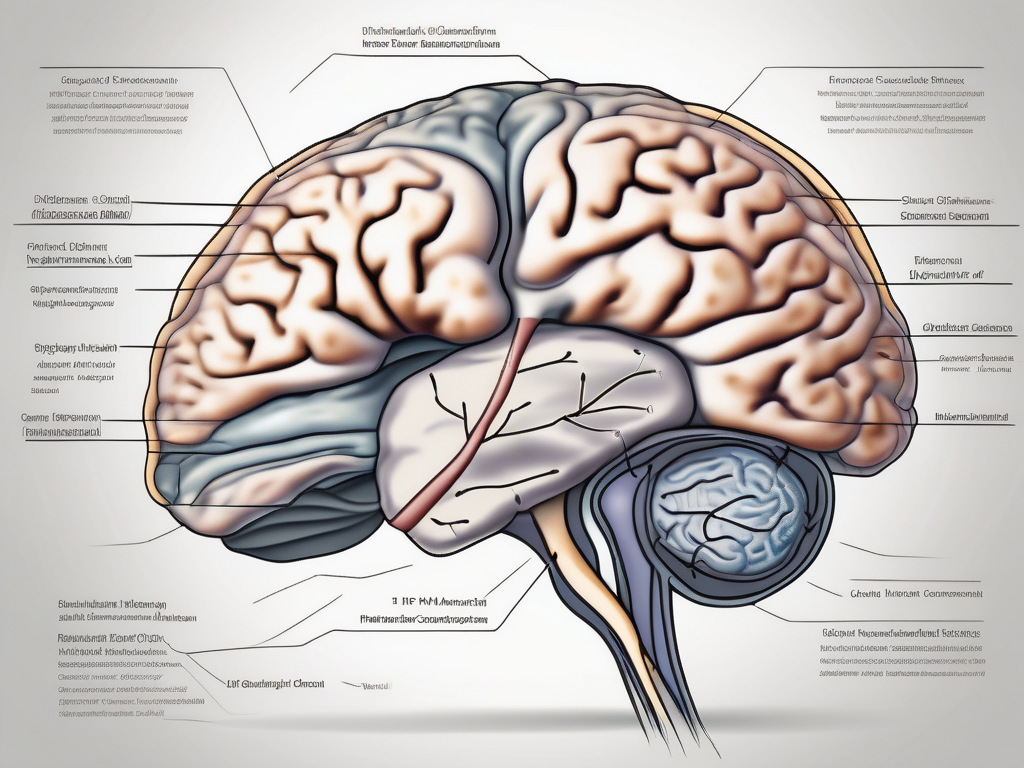
The trochlear nerve, also known as the fourth cranial nerve, plays a crucial role in eye movement. Understanding its anatomy, function, and disorders related to it is essential for gaining insights into the innervation of this intricate nerve. In this article, we will delve into these aspects to provide a comprehensive overview.
Understanding the Trochlear Nerve
The trochlear nerve, also known as the fourth cranial nerve, is a fascinating component of the human nervous system. It is one of the twelve cranial nerves that originate in the brainstem, playing a crucial role in the coordination of eye movements. Let’s delve deeper into the anatomy and function of this remarkable nerve.
Anatomy of the Trochlear Nerve
As mentioned earlier, the trochlear nerve is the smallest of the cranial nerves and possesses a unique pathway. Unlike other cranial nerves that emerge from the brain’s ventral surface, the trochlear nerve exits from the dorsal surface of the midbrain. This distinctive characteristic sets it apart from its counterparts.
The trochlear nerve consists of motor fibers that innervate the superior oblique muscle of the eye. These fibers arise from the trochlear nucleus, a small group of neurons located in the midbrain. Interestingly, these motor fibers decussate, meaning they cross over to the opposite side, before exiting the brainstem. This crossing over of fibers adds an additional layer of complexity to the trochlear nerve’s pathway.
After leaving the brainstem, the trochlear nerve wraps around the brainstem and travels through the subarachnoid space, a fluid-filled space between the brain and the surrounding protective membranes. This journey through the subarachnoid space ensures the nerve’s protection and nourishment before it reaches its final destination.
Upon entering the orbit, the trochlear nerve finally reaches its target, the superior oblique muscle. This muscle, located within the eye socket, plays a crucial role in controlling various eye movements.
Function of the Trochlear Nerve
The primary function of the trochlear nerve is to control the movement of the superior oblique muscle. This muscle’s actions are essential for several crucial eye movements, including depression (downward movement), intorsion (inward rotation), and abduction (outward movement) of the eye.
Imagine looking down at your feet, rotating your eye inward, or moving your eye away from the center of your face. All these movements are made possible by the coordinated action of the trochlear nerve and the superior oblique muscle. Without this precise coordination, our ability to focus on objects and navigate our surroundings would be compromised.
Additionally, the trochlear nerve contributes to maintaining proper alignment of the eyes, ensuring that both eyes work together seamlessly. This alignment is crucial for binocular vision, depth perception, and overall visual acuity.
In conclusion, the trochlear nerve, despite being the smallest cranial nerve, plays a vital role in the complex system that governs our eye movements. Its unique pathway and innervation of the superior oblique muscle make it an intriguing component of the human nervous system. Understanding the trochlear nerve’s anatomy and function provides us with valuable insights into the intricate workings of our visual system.
The Innervation Process of the Trochlear Nerve
When it comes to the innervation process of the trochlear nerve, it involves a complex interaction between various structures and pathways.
The trochlear nerve, also known as the fourth cranial nerve, is responsible for the innervation of the superior oblique muscle, which plays a crucial role in eye movement. This intricate process allows for precise control and coordination of the eyes, enabling us to perform various visual tasks with ease.
Role of the Trochlear Nerve in Eye Movement
The trochlear nerve plays a vital role in eye movement. Specifically, it controls the superior oblique muscle, which allows for downward rotation of the eye, inward tilting, and outward movement, depending on the specific location and state of the muscle.
Imagine you are reading a book. As you move your eyes from one line to another, the trochlear nerve is actively involved in coordinating the downward rotation of your eyes, ensuring that you smoothly transition from one line to the next. This intricate process is essential for efficient reading and comprehension.
Furthermore, the trochlear nerve is also responsible for inward tilting of the eye. This movement is particularly important when focusing on objects that are closer to us. For example, when you are examining a small detail on an object, such as a delicate piece of artwork, the trochlear nerve ensures that your eyes tilt inward, allowing for a clearer and more detailed view.
In addition to downward rotation and inward tilting, the trochlear nerve also facilitates outward movement of the eye. This movement is crucial for tracking objects in our environment. Whether it’s following a flying bird in the sky or keeping an eye on a fast-moving vehicle, the trochlear nerve ensures that our eyes smoothly track the object, allowing us to maintain visual focus and awareness.
The Pathway of the Trochlear Nerve
As the trochlear nerve exits the brainstem, it wraps around the cerebral peduncle and enters the subarachnoid space, traversing the tentorial notch to reach the orbit. Once in the orbit, it innervates the superior oblique muscle, facilitating precise eye movements necessary for tasks such as reading, tracking objects, and maintaining visual fixation.
The pathway of the trochlear nerve is a remarkable journey through the intricate structures of our brain and orbit. Emerging from the dorsal aspect of the midbrain, the nerve wraps around the cerebral peduncle, which is a bundle of nerve fibers connecting the brainstem to the cerebral cortex. This unique route ensures that the trochlear nerve is well-positioned to carry out its important role in eye movement.
After its journey around the cerebral peduncle, the trochlear nerve enters the subarachnoid space, which is a fluid-filled space between the arachnoid mater and pia mater, two of the protective layers surrounding the brain. This space provides a protective cushion for the nerve as it continues its course towards the orbit.
Traversing the tentorial notch, a narrow opening in the tough membrane called the tentorium cerebelli, the trochlear nerve makes its way into the orbit. This notch serves as a passageway, allowing the nerve to reach its final destination and carry out its essential functions.
Once in the orbit, the trochlear nerve innervates the superior oblique muscle, which is responsible for the precise eye movements necessary for various visual tasks. This muscle acts as a pulley, allowing the eye to move smoothly and accurately in different directions. The trochlear nerve’s innervation of the superior oblique muscle ensures that our eyes can perform intricate movements required for activities like reading, tracking objects, and maintaining visual fixation.
In conclusion, the innervation process of the trochlear nerve is a fascinating and intricate system that allows for precise control and coordination of eye movements. From its role in downward rotation, inward tilting, and outward movement of the eye to its unique pathway through the brainstem and orbit, the trochlear nerve plays a crucial role in our visual perception and interaction with the world around us.
Disorders Related to the Trochlear Nerve
Although the trochlear nerve is less commonly affected compared to other cranial nerves, disorders related to its functioning can lead to significant visual disturbances and ocular issues.
The trochlear nerve, also known as the fourth cranial nerve, is responsible for controlling the superior oblique muscle of the eye. This muscle plays a crucial role in eye movement, specifically in downward and inward rotation. When the trochlear nerve is damaged or compressed, it can disrupt the normal functioning of the superior oblique muscle, leading to a variety of symptoms and visual impairments.
Symptoms of Trochlear Nerve Damage
Damage or compression of the trochlear nerve can result in a range of symptoms, including double vision, especially when looking downward. This phenomenon, known as vertical diplopia, occurs because the affected eye is unable to properly align with the unaffected eye, causing a misalignment in the visual field.
In addition to double vision, individuals with trochlear nerve damage may experience difficulty in moving the affected eye. This limitation in eye movement, known as ophthalmoplegia, can make it challenging to focus on objects or track moving targets. The affected eye may also exhibit vertical or torsional misalignment, where it appears higher or lower than the unaffected eye, or rotated in an abnormal position.
Furthermore, some individuals may experience headaches and general eye discomfort as a result of trochlear nerve damage. The strain on the eye muscles and the disruption in normal eye movement can lead to eye strain, tension headaches, and a feeling of overall eye fatigue.
Diagnosis and Treatment of Trochlear Nerve Disorders
Diagnosing disorders related to the trochlear nerve requires careful examination by a healthcare professional, typically an ophthalmologist or a neurologist. They will perform various tests to assess the nerve’s condition and determine the underlying cause of the symptoms.
One of the primary diagnostic tools is a thorough eye examination, which includes assessing visual acuity, eye movement, and alignment. Ocular motility testing, such as the Hess screen test or the Lancaster red-green test, may also be conducted to evaluate the extent of eye muscle weakness or misalignment.
In some cases, imaging scans, such as magnetic resonance imaging (MRI) or computed tomography (CT) scans, may be ordered to visualize the structures of the brain and the cranial nerves. These scans can help identify any structural abnormalities or lesions that may be affecting the trochlear nerve.
Treatment options for trochlear nerve disorders depend on the underlying cause and severity of the condition. In cases where the damage is mild or temporary, conservative approaches such as medication and vision therapy may be recommended. Medications, such as anti-inflammatory drugs or muscle relaxants, can help reduce inflammation and alleviate symptoms. Vision therapy, which involves exercises and techniques to improve eye coordination and muscle strength, may also be beneficial in certain cases.
However, in severe cases where the trochlear nerve damage is significant or irreversible, surgical intervention may be necessary. Surgical procedures, such as trochleoplasty or nerve decompression, aim to repair or release any compression on the trochlear nerve, allowing for improved eye movement and alignment.
It is important for individuals experiencing symptoms related to the trochlear nerve to seek medical advice and consultation with a qualified ophthalmologist or neurologist. Prompt diagnosis and appropriate treatment can help manage the symptoms effectively and prevent further complications.
The Trochlear Nerve in the Wider Nervous System
The trochlear nerve does not work in isolation, but rather functions in harmony with other cranial nerves and the wider nervous system to ensure coordinated eye movements and overall visual functioning.
Interaction of the Trochlear Nerve with Other Cranial Nerves
The trochlear nerve interacts with other cranial nerves, especially the oculomotor nerve (third cranial nerve), to synchronize eye movements. The oculomotor nerve controls the majority of the eye muscles, while the trochlear nerve primarily innervates the superior oblique muscle. Together, they enable precise and coordinated eye movements in different directions.
Eye movements are a complex process that involves the coordinated action of multiple muscles and nerves. The trochlear nerve plays a crucial role in this intricate dance, ensuring that the eyes move smoothly and accurately. It communicates with the oculomotor nerve to transmit signals that control the superior oblique muscle, which is responsible for rotating the eye downwards and outwards. This coordinated movement allows us to focus on objects at different distances and navigate our surroundings with ease.
Furthermore, the trochlear nerve also interacts with the abducens nerve (sixth cranial nerve) to facilitate horizontal eye movements. The abducens nerve controls the lateral rectus muscle, which is responsible for moving the eye laterally. By working together with the abducens nerve, the trochlear nerve ensures that the eyes can scan the environment from side to side, enhancing our visual perception and awareness.
The Trochlear Nerve’s Contribution to Overall Nervous System Functioning
While the primary function of the trochlear nerve lies in eye movement, its contribution extends beyond this specific role. Synchronized eye movements are crucial for depth perception, maintaining balance, and spatial orientation. The trochlear nerve, in conjunction with other cranial nerves and the broader nervous system, aids in these fundamental processes integral to our visual perception and overall functioning.
Depth perception, the ability to perceive the relative distance of objects, relies on the precise coordination of both eyes. The trochlear nerve ensures that the eyes move in perfect synchrony, allowing us to accurately judge distances and perceive the three-dimensional world around us. This is particularly important for activities such as driving, playing sports, and navigating through crowded spaces.
In addition to depth perception, the trochlear nerve also contributes to maintaining balance and spatial orientation. The eyes play a crucial role in our sense of balance, as they provide visual cues that help us orient ourselves in space. By working in harmony with other cranial nerves and the wider nervous system, the trochlear nerve helps us maintain our equilibrium and navigate our surroundings safely.
Moreover, the trochlear nerve’s involvement in spatial orientation allows us to accurately perceive the position of objects in relation to ourselves. This is essential for tasks such as reaching for objects, hand-eye coordination, and spatial reasoning. Without the precise functioning of the trochlear nerve, these everyday activities would be challenging and potentially hazardous.
In conclusion, the innervation of the trochlear nerve is a multifaceted process with significant implications for eye movement, depth perception, balance, and spatial orientation. Understanding the anatomy, function, and disorders related to this nerve provides valuable insights into its intricate role in our daily lives. Should you experience any visual disturbances or suspect trochlear nerve involvement, consulting with a qualified healthcare professional is advised, as they can provide appropriate diagnosis and guide you towards the most suitable treatment options.
