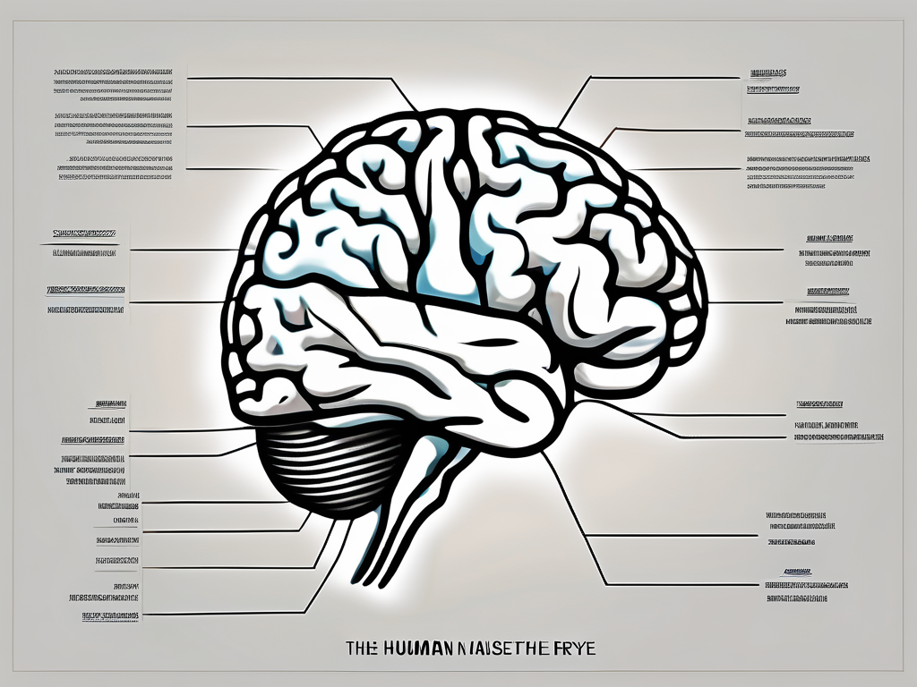
The trochlear nerve, also known as the fourth cranial nerve, plays a crucial role in controlling the movement of certain muscles in our body. Understanding this nerve and its functions can help us appreciate its importance in maintaining optimal body function. In this article, we will delve into the anatomy, function, disorders, treatment, and impact of trochlear nerve damage on daily life.
Understanding the Trochlear Nerve
The trochlear nerve is one of the twelve cranial nerves, emerging from the brainstem at the level of the midbrain. It plays a crucial role in the complex network of nerves that control our vision and eye movements. Specifically, the trochlear nerve is responsible for the movement of the superior oblique muscle of the eye, which allows us to look down and inward.
Anatomy of the Trochlear Nerve
To truly understand the trochlear nerve, it is important to delve into its intricate anatomy. The trochlear nerve originates from the trochlear nucleus, a small structure located in the midbrain. This nucleus serves as the command center for the nerve, coordinating its functions and ensuring smooth communication between the brain and the eye.Interestingly, the trochlear nerve takes a unique pathway within the brainstem. Unlike most other cranial nerves, it undergoes a distinctive decussation, or crossing over, within the brainstem before emerging on the contralateral side of the skull. This crossing over allows for precise coordination of eye movements and ensures that the superior oblique muscle on one side of the head is in sync with the other.
Function of the Trochlear Nerve
The trochlear nerve primarily serves a motor function, providing innervation to the superior oblique muscle. This muscle, as mentioned earlier, plays a crucial role in eye movement. When the trochlear nerve sends signals to the superior oblique muscle, it contracts and causes the eye to rotate downward and inward.This movement is essential for a variety of visual tasks. For instance, when we read a book, the superior oblique muscle helps us look down at the pages and focus on the words. Similarly, when we navigate through a crowded room, the trochlear nerve ensures that our eyes can scan the environment by allowing them to move smoothly and accurately.Moreover, the trochlear nerve also contributes to the rotational movement of the eye. This means that it aids in the precise tracking of objects as they move across our field of vision. Whether it’s following a flying bird or watching a fast-paced tennis match, the trochlear nerve ensures that our eyes can effortlessly track the movement of objects, allowing us to perceive the world around us with clarity and accuracy.In conclusion, the trochlear nerve is a remarkable component of our visual system. Its intricate anatomy and precise control over the superior oblique muscle enable us to perform a wide range of visual tasks, from reading and navigating to tracking moving objects. Without the trochlear nerve, our ability to move our eyes in a coordinated manner would be greatly compromised.
Muscles Controlled by the Trochlear Nerve
The trochlear nerve primarily controls the superior oblique muscle, which is responsible for various essential eye movements. Let’s explore this muscle and the role of the trochlear nerve in eye movement.
Superior Oblique Muscle
The superior oblique muscle is one of the six extraocular muscles responsible for eye movement. It originates from the common tendinous ring located at the back of the eye socket. This muscle loops through a cushion-like tendon, called the trochlea, before inserting into the upper, outer surface of the eyeball. The superior oblique muscle is unique in its path, as it is the only muscle that passes through a pulley-like structure. This pulley mechanism allows the muscle to change its direction of pull, enabling it to perform its specific functions.The primary function of the superior oblique muscle is to rotate the eye in a downward and outward direction. This movement is known as depression and abduction. When the superior oblique muscle contracts, it pulls the eye downward and away from the midline, allowing us to look down and to the side. This movement is crucial for tasks such as reading, driving, and navigating our surroundings.In addition to its role in eye movement, the superior oblique muscle also plays a vital role in maintaining visual alignment. It works in conjunction with the other eye muscles to ensure that both eyes are properly aligned and focused on the same point. This coordination is essential for binocular vision, depth perception, and overall visual acuity.
Role of Trochlear Nerve in Eye Movement
The trochlear nerve, also known as the fourth cranial nerve, innervates the superior oblique muscle, playing a vital role in eye movement coordination. It originates from the midbrain and has the longest intracranial course of all the cranial nerves. The trochlear nerve exits the brainstem dorsally and crosses over to the opposite side, controlling the contralateral superior oblique muscle.Dysfunction of the trochlear nerve can lead to impaired eye movement, causing double vision, difficulties in focusing, and visual disturbances. Trochlear nerve palsy, a condition characterized by weakness or paralysis of the superior oblique muscle, can result from various causes such as trauma, infection, or vascular disorders. This condition can significantly impact a person’s ability to perform daily activities that require precise eye movements, such as reading, driving, or playing sports.In conclusion, the trochlear nerve and the superior oblique muscle work together to ensure proper eye movement and visual alignment. Understanding the anatomy and function of these structures is crucial in diagnosing and managing conditions that affect eye movement coordination. Further research and advancements in this field will continue to enhance our understanding of the complex mechanisms underlying eye movement and contribute to the development of effective treatments for related disorders.
Disorders Related to the Trochlear Nerve
Various disorders can affect the trochlear nerve, resulting in functional impairment. Let’s explore the causes, symptoms, and diagnosis of trochlear nerve disorders.
Causes of Trochlear Nerve Palsy
Trochlear nerve palsy, or dysfunction, may occur due to various factors. One common cause is trauma, such as a direct blow to the head or an injury to the eye socket. In some cases, congenital anomalies, which are present at birth, can affect the trochlear nerve. These anomalies can include abnormal development of the nerve or its surrounding structures.Infections can also lead to trochlear nerve disorders. Conditions like meningitis or sinusitis can cause inflammation in the area, affecting the function of the nerve. Additionally, tumors, both benign and malignant, can put pressure on the trochlear nerve, leading to dysfunction.Vascular diseases, such as aneurysms or arteriovenous malformations, can disrupt the blood supply to the trochlear nerve, impairing its function. Lastly, neurological conditions like multiple sclerosis, which affects the central nervous system, can also result in trochlear nerve palsy. The exact cause of trochlear nerve disorders can vary among individuals, necessitating a comprehensive evaluation by a medical professional.
Symptoms and Diagnosis of Trochlear Nerve Disorders
Trochlear nerve disorders can manifest with various symptoms, which can vary depending on the severity of the dysfunction. One common symptom is diplopia, or double vision. This occurs because the affected eye is unable to move properly, resulting in the brain receiving conflicting visual information from both eyes.Individuals with trochlear nerve disorders may also experience difficulty looking downwards. This can make activities such as reading or walking down stairs challenging. To compensate for the impaired downward gaze, some individuals may tilt their head in an attempt to improve their vision.Eye misalignment is another common symptom of trochlear nerve disorders. The affected eye may appear higher or lower than the unaffected eye, leading to an uneven gaze. Problems with depth perception can also occur, making it difficult to accurately judge distances.Proper diagnosis of trochlear nerve disorders involves a thorough medical history review and physical examination. The medical professional will inquire about the onset and progression of symptoms, as well as any relevant medical conditions or previous injuries. During the physical examination, the doctor may assess eye movements, looking for any abnormalities or limitations.In some cases, additional diagnostic tests may be necessary to confirm the diagnosis. Imaging studies, such as magnetic resonance imaging (MRI) or computed tomography (CT) scans, can provide detailed images of the trochlear nerve and surrounding structures. Electromyography (EMG) may also be performed to assess the electrical activity of the muscles controlled by the trochlear nerve.In conclusion, trochlear nerve disorders can have various causes, including trauma, congenital anomalies, infections, tumors, vascular diseases, and neurological conditions. The symptoms of these disorders can range from diplopia and difficulty looking downwards to eye misalignment and problems with depth perception. Proper diagnosis involves a comprehensive evaluation, including a medical history review, physical examination, and potentially additional diagnostic tests.
Treatment and Management of Trochlear Nerve Disorders
Treatment for trochlear nerve disorders aims to alleviate symptoms and improve functionality, depending on the underlying cause. Non-surgical and surgical interventions are available, but treatment plans should always be determined by a healthcare professional.
Non-Surgical Treatments
Non-surgical approaches may include the use of prism glasses to address double vision or eye patches to improve visual alignment. These interventions can help patients with trochlear nerve disorders regain a sense of visual stability and reduce the discomfort associated with misaligned eyes.
In addition to optical aids, physical therapy exercises can also be beneficial in strengthening the affected eye muscles and improving eye coordination. These exercises may include eye tracking exercises, focusing exercises, and eye muscle strengthening exercises. Physical therapists specializing in ophthalmic rehabilitation can guide patients through these exercises, ensuring proper technique and progression.
Furthermore, non-surgical treatments may involve the use of medications to manage symptoms such as pain, inflammation, or muscle spasms. These medications can provide temporary relief and improve the overall comfort of individuals with trochlear nerve disorders.
Surgical Interventions
In some cases, surgical intervention may be necessary to correct underlying anatomical abnormalities or reposition the attached tendons to improve eye movement. Surgical options are tailored to each individual’s specific needs, and consulting with a specialized ophthalmologist is critical.
One surgical procedure commonly used to treat trochlear nerve disorders is trochleoplasty. This procedure involves reshaping the trochlea, the bony structure that the trochlear nerve passes through, to improve the alignment and movement of the affected eye. Trochleoplasty is often performed in conjunction with other surgical techniques, such as tendon repositioning or muscle tightening, to achieve optimal results.
Another surgical option is tenotomy, which involves the release or lengthening of the tight or shortened tendons that may be causing restricted eye movement. This procedure allows for greater flexibility and improved range of motion in the affected eye.
It is important to note that surgical interventions for trochlear nerve disorders carry risks and potential complications. These can include infection, bleeding, scarring, and changes in vision. Therefore, it is crucial for patients to have a thorough discussion with their ophthalmologist to fully understand the potential benefits and risks associated with any surgical procedure.
Post-surgical care and rehabilitation are also essential for optimal outcomes. Following surgery, patients may be prescribed eye exercises and physical therapy to aid in the recovery process and maximize the benefits of the surgical intervention.
In conclusion, the treatment and management of trochlear nerve disorders involve a range of non-surgical and surgical interventions. These treatments aim to alleviate symptoms, improve functionality, and enhance the overall quality of life for individuals affected by trochlear nerve disorders. Consulting with a healthcare professional specializing in ophthalmology is crucial in developing an appropriate treatment plan tailored to each individual’s unique needs.
The Impact of Trochlear Nerve Damage on Daily Life
Trochlear nerve damage can significantly impact an individual’s daily life, particularly in activities that require precise eye movement and coordination. Let’s explore the challenges faced and coping mechanisms that can be employed.
Vision Impairment and Adaptation
Individuals with trochlear nerve damage may experience vision impairment, such as difficulties in reading, driving, or participating in sports. This can be incredibly frustrating and can have a profound impact on one’s independence and overall quality of life.
Adapting to these challenges often involves visual exercises, assistive devices, and lifestyle modifications to maximize visual function. Visual exercises, such as eye tracking and coordination exercises, can help strengthen the remaining muscles and improve eye movement control. Assistive devices, such as magnifiers or special glasses, can aid in reading and other visual tasks. Lifestyle modifications, such as adjusting lighting conditions or using larger fonts, can also make a significant difference in daily activities.
Furthermore, individuals with trochlear nerve damage may need to learn new strategies to compensate for their vision impairment. For example, they may develop techniques to scan their environment more efficiently or rely more on auditory cues to navigate their surroundings. These adaptive strategies can help individuals regain a sense of independence and continue participating in activities they enjoy.
Coping with Trochlear Nerve Disorders
Coping with trochlear nerve disorders can be challenging, both physically and emotionally. It is essential for individuals to seek support from healthcare professionals who specialize in neurology or ophthalmology. These experts can provide a proper diagnosis, recommend appropriate treatment options, and offer guidance on managing the condition.
In addition to medical support, joining support groups can be immensely beneficial. Connecting with others who are going through similar experiences can provide a sense of understanding, validation, and emotional support. Support groups can also be a valuable source of information, as members share their coping strategies and experiences with different treatment approaches.
Utilizing resources available to enhance quality of life is also crucial. This may include accessing assistive technology, such as screen readers or speech recognition software, to facilitate communication and access to information. Occupational therapists can provide valuable guidance on adapting daily activities and recommending assistive devices that can make tasks easier to manage.
It is important to remember that each person’s experience with trochlear nerve damage is unique. While some individuals may experience mild symptoms that are easily managed, others may face more significant challenges. Consulting with medical experts is crucial for appropriate guidance and management tailored to individual needs.
In conclusion, the trochlear nerve plays a crucial role in controlling specific muscles, primarily the superior oblique muscle, responsible for eye movement. Understanding the anatomy, function, disorders, treatment options, and impact of trochlear nerve damage can empower individuals to take proactive steps in managing their visual health. However, it is crucial to consult with medical professionals for proper diagnosis, treatment planning, and ongoing care.
