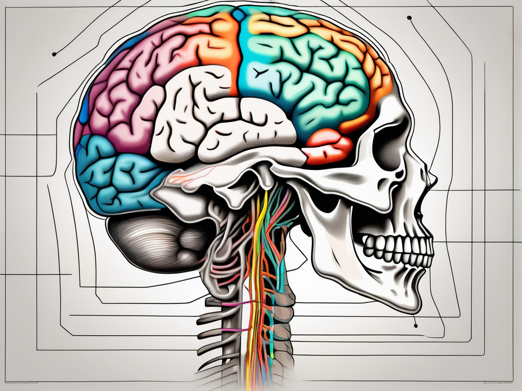
The trochlear nerve is an important cranial nerve that plays a crucial role in the fine movements of the eye. It is the fourth cranial nerve and can be found in the brainstem. To better understand this nerve and its significance, let’s delve into its definition, function, anatomy, pathway, clinical significance, and the treatment options available for any associated disorders.
Understanding the Trochlear Nerve
The trochlear nerve, also known as cranial nerve IV, is a fascinating component of the human body’s intricate nervous system. This nerve plays a crucial role in facilitating the movement of the superior oblique muscle of the eye, which is responsible for rotating the eye upwards and inwards. By understanding the anatomy and function of the trochlear nerve, we can gain a deeper appreciation for the complexity and precision of our visual system.
Definition and Function of the Trochlear Nerve
The trochlear nerve, as mentioned earlier, is responsible for the movement of the superior oblique muscle of the eye. This muscle aids in the complex process of eye movement, allowing us to focus on objects at different distances and angles. The trochlear nerve arises from the dorsal aspect of the mesencephalon, also known as the midbrain, and connects the brain to the muscle it innervates. This connection enables precise eye movements, ensuring that our vision remains clear and accurate.
Imagine the trochlear nerve as a vital communication pathway between the brain and the superior oblique muscle. Without this nerve, our ability to move our eyes in a coordinated manner would be severely compromised. Simple tasks such as reading, driving, or even following the flight of a bird would become challenging and potentially impossible.
The Anatomy of the Trochlear Nerve
Now, let’s delve into the fascinating anatomy of the trochlear nerve. This nerve emerges from the posterior surface of the midbrain, making it unique among the cranial nerves. Unlike its counterparts, the trochlear nerve exits the brainstem dorsally, rather than ventrally. This distinctive pathway sets it apart from the other cranial nerves and contributes to its vulnerability to injury or damage.
As the trochlear nerve emerges from the midbrain, it takes a remarkable journey, wrapping around the midbrain in a loop-like structure. This looping pathway is known as the trochlear loop. This anatomical peculiarity adds another layer of complexity to the trochlear nerve’s function and vulnerability. The loop-like structure serves as a protective mechanism, shielding the nerve from potential damage as it makes its way towards its destination.
Eventually, the trochlear nerve exits the skull through a small opening called the superior orbital fissure. This exit point allows the nerve to reach the superior oblique muscle of the eye, where it can carry out its essential function of facilitating precise eye movements. However, the location of the superior orbital fissure also exposes the trochlear nerve to potential trauma or compression, which can lead to various visual impairments.
Understanding the intricate anatomy of the trochlear nerve highlights the delicate balance required for optimal eye function. The slightest disruption or damage to this nerve can have significant implications for our vision and overall quality of life. It serves as a reminder of the remarkable complexity and interconnectedness of the human body.
The Pathway of the Trochlear Nerve
Origin and Termination of the Trochlear Nerve
The trochlear nerve, also known as the fourth cranial nerve, plays a crucial role in eye movement. It originates from the trochlear nucleus, a small group of neurons located in the midbrain just below the cerebral aqueduct. This nucleus is responsible for controlling the superior oblique muscle, one of the extraocular muscles that helps with eye movement.
Within the trochlear nucleus, the axons of the trochlear nerve arise and then decussate, or cross over, within the brainstem. This decussation is a unique feature of the trochlear nerve, as it is the only cranial nerve to do so. After crossing over, the axons exit the brainstem and continue their journey towards their target muscle.
The trochlear nerve exits the brain through the posterior aspect of the midbrain, specifically through the superior medullary velum. From there, it enters the subarachnoid space, which is the space between the arachnoid mater and the pia mater, two of the meninges that protect the brain and spinal cord.
As the trochlear nerve travels through the subarachnoid space, it takes a complex course, navigating around various structures within the brain. It passes ventrally, or towards the front, and wraps around the midbrain, forming the trochlear nucleus. This unique path allows the trochlear nerve to reach its target muscle from a specific angle, optimizing its function.
The Course of the Trochlear Nerve in the Brain
Continuing its journey, the trochlear nerve winds around the midbrain, carefully avoiding other cranial nerves and important structures. This intricate pathway ensures that the trochlear nerve remains protected and can function optimally.
As the trochlear nerve reaches the inferior surface of the midbrain, it approaches the superior orbital fissure, a narrow opening located in the sphenoid bone. This fissure serves as a passageway for several structures, including the trochlear nerve.
Finally, the trochlear nerve exits the skull through the superior orbital fissure and enters the orbit, or eye socket. Here, it reaches its target muscle—the superior oblique muscle of the eye. The superior oblique muscle plays a crucial role in eye movement, allowing for downward and inward rotation of the eye.
In conclusion, the pathway of the trochlear nerve is a fascinating journey through the brain and skull. From its origin in the midbrain to its termination in the superior oblique muscle, this cranial nerve takes a complex and intricate course, ensuring precise control of eye movement. Understanding the pathway of the trochlear nerve is essential for comprehending the mechanisms behind eye movement and the intricate connections within the brain.
Clinical Significance of the Trochlear Nerve
The trochlear nerve, also known as the fourth cranial nerve, plays a crucial role in eye movement. It is responsible for innervating the superior oblique muscle, which helps control the downward and inward movement of the eye. Despite being relatively protected within the brain, the trochlear nerve can still be affected by various conditions, leading to significant clinical implications.
Common Disorders Involving the Trochlear Nerve
One of the most common disorders associated with the trochlear nerve is trochlear nerve palsy, also known as fourth nerve palsy. This condition is characterized by weakness or paralysis of the superior oblique muscle, resulting in a range of visual impairments. Individuals with trochlear nerve palsy often experience vertical or torsional double vision, where objects appear to be either stacked on top of each other or tilted.
The impact of trochlear nerve palsy on a person’s daily life and well-being cannot be overstated. Simple tasks such as reading, driving, or even walking can become challenging and frustrating. The constant struggle to align visual images can lead to eye strain, headaches, and a decreased quality of life.
While the exact cause of trochlear nerve palsy can vary, it is often the result of trauma, such as head injuries or fractures involving the orbit. In some cases, the condition may be congenital, meaning it is present at birth. Other potential causes include infections, tumors, or vascular abnormalities affecting the nerve.
Diagnostic Tests for Trochlear Nerve Damage
When trochlear nerve dysfunction is suspected, doctors employ various diagnostic tests to assess the extent of the damage and identify the underlying cause. A comprehensive eye examination is typically the first step in evaluating eye movements and potential impairments. This examination may involve assessing visual acuity, pupillary reflexes, and extraocular muscle function.
Specifically, the function of the cranial nerves, including the trochlear nerve, is evaluated through tests such as the Hirschberg test, which assesses the alignment of the eyes, and the cover-uncover test, which detects any misalignment or deviation. These tests help determine the presence and severity of trochlear nerve palsy.
In addition to a clinical examination, imaging tests may be ordered to identify any structural abnormalities or lesions affecting the trochlear nerve. Magnetic resonance imaging (MRI) is commonly used to visualize the brain and surrounding structures in detail. It can help detect tumors, vascular malformations, or other pathologies that may be compressing or damaging the trochlear nerve.
Overall, the clinical significance of the trochlear nerve cannot be overlooked. Its role in eye movement and the potential disorders associated with it highlight the importance of early diagnosis and appropriate management. By understanding the clinical implications and utilizing diagnostic tests, healthcare professionals can provide targeted interventions and improve the quality of life for individuals affected by trochlear nerve dysfunction.
Treatment and Management of Trochlear Nerve Disorders
Trochlear nerve disorders can cause a range of symptoms, including double vision and difficulty moving the affected eye. The treatment of these disorders depends on the underlying cause and the severity of the condition. In some cases, non-surgical approaches may be sufficient to manage symptoms, while in other cases, surgical interventions may be necessary.
Non-Surgical Treatment Options
Non-surgical treatment options for trochlear nerve disorders aim to alleviate symptoms and improve the functionality of the affected eye muscle. One common non-surgical approach is the use of prism glasses. These specialized glasses can help correct double vision by bending light in a way that allows the eyes to align properly. By wearing prism glasses, individuals with trochlear nerve disorders can experience improved vision and reduced eye strain.
In addition to prism glasses, eye exercises may also be prescribed as a non-surgical treatment option. These exercises are designed to strengthen the affected eye muscle and improve its ability to move the eye in the correct direction. Eye exercises can be tailored to the individual’s specific needs and can be performed under the guidance of a healthcare professional.
It is important to note that the effectiveness of non-surgical treatment options may vary depending on the severity of the trochlear nerve disorder. In some cases, these approaches may provide significant relief, while in others, they may only offer temporary improvement. Therefore, it is crucial to consult with a healthcare professional to determine the most appropriate treatment plan.
Surgical Interventions for Trochlear Nerve Damage
In cases where conservative measures fail to adequately address the symptoms or if the trochlear nerve is severely damaged, surgical interventions may be considered. These surgeries aim to correct the alignment of the eyes and improve the functionality of the superior oblique muscle, which is responsible for the movement of the affected eye.
There are different surgical techniques that can be used to treat trochlear nerve damage. One common procedure is trochleoplasty, which involves reshaping the groove in the eye socket where the trochlear nerve passes through. By modifying the shape of the groove, the movement of the superior oblique muscle can be improved, leading to better eye alignment and reduced symptoms.
In some cases, additional procedures may be performed in conjunction with trochleoplasty. These can include tendon transfers, where a tendon from another part of the body is used to strengthen the affected eye muscle, or muscle resection, where a portion of the muscle is removed to improve its functionality.
It is important to emphasize that the decision to undergo surgery for trochlear nerve disorders should only be made after careful consultation with a qualified medical specialist. The potential risks and benefits of the procedure should be thoroughly discussed, and the individual’s overall health and specific circumstances should be taken into consideration.
In conclusion, the treatment and management of trochlear nerve disorders require a comprehensive approach that takes into account the underlying cause and the severity of the condition. Non-surgical options, such as prism glasses and eye exercises, can provide relief for some individuals, while others may require surgical interventions to improve eye alignment and functionality. It is crucial to work closely with a healthcare professional to determine the most appropriate treatment plan and to ensure the best possible outcome for those affected by trochlear nerve disorders.
Prevention and Prognosis of Trochlear Nerve Disorders
Preventive Measures for Trochlear Nerve Health
While trochlear nerve disorders can arise from various causes, adopting a healthy lifestyle and taking preventive measures can contribute to overall eye health. Regular eye examinations, maintaining a balanced diet, protecting the eyes from trauma, and avoiding excessive strain on the eyes can all help in reducing the risk of trochlear nerve dysfunction.
Regular eye examinations are essential for maintaining optimal eye health. During these examinations, eye care professionals can assess the condition of the trochlear nerve and detect any early signs of dysfunction. They can also provide guidance on how to protect the eyes and prevent potential damage to the nerve.
A balanced diet rich in nutrients is crucial for the health of the trochlear nerve. Certain vitamins and minerals, such as vitamin A, vitamin C, and omega-3 fatty acids, play a vital role in maintaining the integrity of the nerves and supporting overall eye health. Including foods like carrots, leafy greens, citrus fruits, and fatty fish in your diet can help provide these essential nutrients.
Protecting the eyes from trauma is another important preventive measure. Wearing appropriate protective eyewear during activities that pose a risk of eye injury, such as sports or certain occupations, can help prevent damage to the trochlear nerve. Additionally, avoiding activities that put excessive strain on the eyes, such as prolonged screen time or reading in poor lighting conditions, can help reduce the risk of nerve dysfunction.
Prognosis and Recovery from Trochlear Nerve Disorders
The prognosis for trochlear nerve disorders varies depending on the specific condition and individual factors. In many cases, with appropriate treatment and management, individuals can experience significant improvement in their symptoms. However, early diagnosis and intervention are crucial for a better prognosis. It is essential to consult with a healthcare professional to receive an accurate diagnosis and appropriate treatment plan.
Early diagnosis plays a crucial role in the prognosis of trochlear nerve disorders. Identifying the underlying cause of the dysfunction and implementing targeted treatment strategies can help alleviate symptoms and prevent further damage to the nerve. Healthcare professionals, such as neurologists or ophthalmologists, may perform various diagnostic tests, such as imaging scans or nerve conduction studies, to determine the extent of the nerve damage and develop an effective treatment plan.
Treatment options for trochlear nerve disorders may include medication, physical therapy, or in severe cases, surgical intervention. Medications, such as pain relievers or anti-inflammatory drugs, can help manage symptoms and reduce inflammation around the nerve. Physical therapy techniques, such as eye exercises or eye muscle strengthening exercises, may be recommended to improve eye coordination and control. In some cases, surgical procedures, such as decompression surgery or nerve repair, may be necessary to restore normal nerve function.
Recovery from trochlear nerve disorders can vary from person to person. Factors such as the severity of the nerve damage, individual response to treatment, and adherence to the recommended therapy plan can all influence the recovery process. It is important to follow the healthcare professional’s guidance and attend regular follow-up appointments to monitor progress and make any necessary adjustments to the treatment plan.
In conclusion, the trochlear nerve, situated in the midbrain, plays a vital role in the fine movements of the eye. Understanding its anatomy, function, and clinical significance are essential for recognizing and managing trochlear nerve disorders. If you suspect any issues related to this nerve or experience visual disturbances, it is advisable to consult with a healthcare professional who can provide an accurate diagnosis and guide you towards appropriate treatment options.
