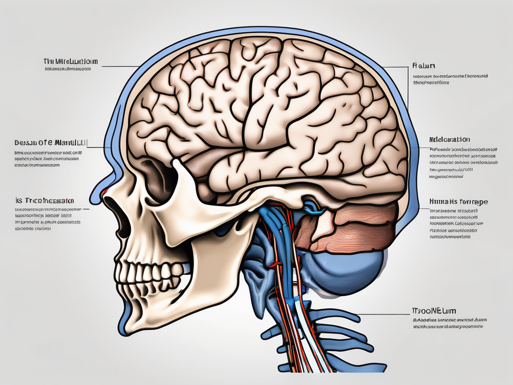
The trochlear nerve is a vital component of the human nervous system, responsible for the movement of one of the six extraocular muscles. Understanding the intricate anatomy and function of this nerve is essential in comprehending its pathway, crossing point, and exit point. Disorders associated with the trochlear nerve can have significant implications on a person’s vision and overall quality of life. In this article, we delve into the fascinating world of the trochlear nerve, exploring its anatomy, functions, pathway, and various related disorders. By shedding light on these aspects, we hope to provide a comprehensive understanding of where exactly the trochlear nerve crosses and exits the human body.
Understanding the Trochlear Nerve
The trochlear nerve, also known as the fourth cranial nerve or CN IV, is a motor nerve that primarily controls the superior oblique muscle of the eye. This muscle is responsible for various eye movements, including depression, intorsion, and abduction. The trochlear nerve emerges from the posterior midbrain, specifically the trochlear nucleus. It is the only cranial nerve to decussate (cross over) inside the brain, a unique characteristic that plays a significant role in its functioning.
Anatomy of the Trochlear Nerve
The trochlear nerve has the longest intracranial course of all the cranial nerves. It originates from the superior colliculus in the posterior dorsal region of the midbrain. Following its emergence, the trochlear nerve wraps around the midbrain’s lateral surface, coursing upward and backward towards the cavernous sinus. A distinct feature of the trochlear nerve is that it innervates a single muscle, the superior oblique, making it anatomically and functionally unique among the cranial nerves.
As the trochlear nerve travels along its intricate pathway, it passes through various structures within the brain, including the red nucleus, which is involved in motor coordination. This close proximity to other important brain regions highlights the interconnectedness of the nervous system and the complex nature of eye movement control.
Additionally, the trochlear nerve’s long intracranial course presents a potential vulnerability. Its extensive journey leaves it susceptible to damage from trauma or compression, which can result in trochlear nerve palsy. This condition leads to weakness or paralysis of the superior oblique muscle, causing a range of visual disturbances and challenges in daily activities.
Functions of the Trochlear Nerve
The trochlear nerve’s primary function is to facilitate the movement of the superior oblique muscle. Working in conjunction with the other extraocular muscles and cranial nerves responsible for eye movement, the trochlear nerve allows for coordinated and precise control of eye position and orientation. Dysfunction of the trochlear nerve can lead to diplopia (double vision), vertical misalignment, and difficulty in focusing properly, resulting in a range of visual disturbances.
Moreover, the trochlear nerve plays a crucial role in depth perception. By controlling the superior oblique muscle, it helps to create the necessary tension and alignment in the eyes to accurately perceive depth and distance. This ability is essential for activities such as judging the distance of an object, catching a ball, or driving a car.
Furthermore, the trochlear nerve contributes to the stabilization of eye movements during head movements. It works in tandem with the vestibulo-ocular reflex, a mechanism that allows the eyes to move in the opposite direction to head movements, ensuring a stable and clear visual field. This coordination is vital for maintaining visual acuity and preventing blurring or disorientation during rapid head movements.
Interestingly, the trochlear nerve’s unique decussation within the brain allows for contralateral control of the superior oblique muscle. This means that the left trochlear nerve controls the right superior oblique muscle, and vice versa. This crossed arrangement ensures the precise coordination and synchronization of eye movements, contributing to binocular vision and the ability to perceive depth accurately.
In conclusion, the trochlear nerve is a remarkable cranial nerve that plays a crucial role in eye movement control and visual perception. Its intricate anatomy, long intracranial course, and unique decussation within the brain highlight its significance in maintaining proper eye alignment, depth perception, and stability during head movements. Understanding the trochlear nerve’s functions and characteristics is essential for comprehending the complexities of the visual system and the intricate interplay between the nervous system and ocular motor control.
Pathway of the Trochlear Nerve
The pathway of the trochlear nerve encompasses its origin and course, as well as the crossing point and exit point. Understanding these aspects is crucial to grasp the nerve’s trajectory within the human body.
Origin and Course of the Trochlear Nerve
The trochlear nerve arises in the posterior midbrain, specifically from the trochlear nucleus. This small nucleus is located dorsally in the midbrain, near the cerebral aqueduct. From its origin, the delicate nerve fiber bundles decussate within the brainstem, meaning that the fibers from one side cross over to the opposite side. This decussation occurs at the level of the superior medullary velum, which is a thin membrane-like structure in the midbrain.
After decussating, the trochlear nerve fibers exit the ventral surface of the midbrain. While other cranial nerves exit the brainstem laterally, the trochlear nerve takes a unique posterior exit, beginning its downward and backward journey. As the nerve fibers leave the midbrain, they form a compact bundle that travels through the subarachnoid space, which is the space between the arachnoid mater and the pia mater, two of the layers that surround the brain.
As the trochlear nerve continues its course, it wraps around the cerebral peduncles, which are large bundles of nerve fibers that connect the midbrain to the rest of the brain. This curved trajectory allows the nerve to avoid other structures in the brain, ensuring its proper functioning and avoiding any potential damage.
Crossing Point of the Trochlear Nerve
Inside the brain, the trochlear nerve decussates, or crosses over, at the level of the superior medullary velum. This crossing point is crucial for the proper innervation of the superior oblique muscle, one of the extraocular muscles responsible for eye movement. The crossed fibers of the trochlear nerve then continue their journey, following a curved and elongated course along the brainstem.
As the trochlear nerve fibers travel along the brainstem, they pass through several important structures. They navigate through the tectum, which is the dorsal part of the midbrain responsible for sensory processing. The nerve fibers also pass near the red nucleus, a structure involved in motor coordination. This close proximity to other important brain structures highlights the intricate nature of the trochlear nerve’s pathway.
Eventually, the trochlear nerve fibers reach the superior orbital fissure, a narrow opening located in the posterior part of the orbit, or eye socket. This exit point allows the nerve to leave the brain and enter the orbit, where it innervates the superior oblique muscle. The superior oblique muscle plays a crucial role in eye movement, specifically in rotating the eye downward and outward.
In summary, the pathway of the trochlear nerve involves its origin in the trochlear nucleus of the midbrain, decussation at the superior medullary velum, and a curved course along the brainstem. This intricate trajectory ensures proper innervation of the superior oblique muscle, allowing for precise eye movements. Understanding the pathway of the trochlear nerve provides valuable insights into the complex network of nerves within the human body.
Exit Point of the Trochlear Nerve
Identifying the exit point of the trochlear nerve is essential in determining its anatomical location and its relationship with adjacent structures. The trochlear nerve’s exit point lies at the anterior surface of the brainstem, near the junction between the midbrain and pons.
The trochlear nerve, also known as the fourth cranial nerve, is responsible for the innervation of the superior oblique muscle of the eye. This muscle plays a crucial role in eye movement, specifically in rotating the eye downward and outward. Understanding the exit point of the trochlear nerve is vital for comprehending its function and potential clinical implications.
Identifying the Exit Point
The exit point of the trochlear nerve is situated below the inferior colliculus and above the pontomesencephalic junction. Accurate localization of this exit point helps clinicians and surgeons evaluate possible compression or damage to the trochlear nerve that may contribute to visual impairments.
When the trochlear nerve exits the brainstem, it passes through the cavernous sinus, a venous channel located on each side of the sella turcica. The cavernous sinus contains important structures such as the internal carotid artery, oculomotor nerve, and abducens nerve. The proximity of the trochlear nerve’s exit point to these structures highlights the need for precise identification to avoid potential complications during surgical procedures.
Significance of the Exit Point
The exit point of the trochlear nerve holds great clinical significance. It is a crucial landmark for surgical interventions and diagnostic imaging tools. By precisely identifying the exit point, healthcare professionals can devise appropriate treatment plans for patients experiencing trochlear nerve-related disorders.
Lesions or injuries to the trochlear nerve can result in a condition called trochlear nerve palsy, which leads to weakness or paralysis of the superior oblique muscle. This can cause vertical diplopia (double vision) and difficulty in downward and outward eye movements. Understanding the exit point of the trochlear nerve aids in diagnosing and managing such conditions effectively.
Furthermore, the exit point of the trochlear nerve serves as a reference point for neurosurgeons when performing surgical procedures in the region. It allows for precise localization and avoidance of damage to the nerve during interventions, ensuring optimal patient outcomes.
Disorders Related to the Trochlear Nerve
Despite its significance, the trochlear nerve is susceptible to various disorders that can have a profound impact on eye movement and visual acuity.
The trochlear nerve, also known as the fourth cranial nerve, plays a crucial role in the control of eye movement. It is responsible for innervating the superior oblique muscle, which helps rotate the eye downward and outward. Any disruption or damage to this nerve can lead to a range of disorders that affect the normal functioning of the eye.
Common Trochlear Nerve Disorders
Pathologies affecting the trochlear nerve can range from congenital malformations to acquired conditions. Common disorders include trochlear nerve palsy, hypertropia, and trochleitis. Trochlear nerve palsy, also known as fourth nerve palsy, is a condition characterized by weakness or paralysis of the superior oblique muscle. This can result in vertical misalignment of the eyes, leading to diplopia (double vision) in affected individuals.
Hypertropia, on the other hand, refers to an abnormal upward deviation of one eye in relation to the other. It can be caused by various factors, including trochlear nerve dysfunction. Individuals with hypertropia often experience difficulties in maintaining binocular vision, which can significantly impact their daily activities.
Trochleitis is a rare condition characterized by inflammation of the trochlear nerve. It can be caused by infections, autoimmune disorders, or trauma. Symptoms of trochleitis may include eye pain, redness, and limited eye movement. Prompt diagnosis and treatment are crucial to prevent further complications and alleviate the associated symptoms.
Symptoms and Diagnosis of Trochlear Nerve Disorders
Symptoms of trochlear nerve disorders may include double vision, difficulty looking downwards, and tilting of the head to avoid diplopia. These symptoms can vary in severity depending on the underlying cause and the extent of nerve damage. It is important to note that these symptoms can also be indicative of other eye-related conditions, highlighting the need for a comprehensive evaluation by a medical professional.
Accurate diagnosis of trochlear nerve disorders usually involves a thorough assessment of the patient’s medical history, a comprehensive ophthalmological examination, and in some cases, neuroimaging studies. The medical history assessment helps identify any underlying conditions or previous trauma that may have contributed to the development of the disorder. The ophthalmological examination includes tests to assess eye movement, visual acuity, and binocular vision. Neuroimaging studies, such as magnetic resonance imaging (MRI), may be recommended to visualize the trochlear nerve and identify any structural abnormalities or lesions.
It is essential to consult with a medical professional for an accurate diagnosis and appropriate treatment. Early detection and intervention can significantly improve the prognosis and quality of life for individuals affected by trochlear nerve disorders.
Treatment and Management of Trochlear Nerve Disorders
When it comes to treating trochlear nerve disorders, there are several approaches that healthcare professionals can take to address the condition effectively. These approaches include medical treatments, surgical interventions, and rehabilitation options.
Medical Treatments
In some cases, trochlear nerve disorders may be managed through conservative measures. One such measure is occlusion therapy, which involves covering one eye to encourage the affected eye to work harder and improve its muscle control. Another conservative approach is the use of prism glasses, which can help correct double vision caused by trochlear nerve disorders. Additionally, pharmacological interventions may be prescribed to address the underlying cause of the disorder. These medications can help alleviate symptoms and promote healing. The specific treatment plan will depend on the individual patient’s condition and the severity of their symptoms.
Surgical Interventions
In more severe cases or when conservative measures prove ineffective, surgical interventions may be necessary to provide long-term relief. These procedures aim to address nerve compression, correct muscle imbalances, and restore proper alignment and function of the eye. Surgical techniques vary depending on the nature and extent of the trochlear nerve disorder. It is crucial to consult with an experienced ophthalmologist or neurosurgeon for an accurate assessment and tailored treatment plan. They will consider factors such as the patient’s overall health, the specific symptoms experienced, and the potential risks and benefits of surgery.
Rehabilitation and Therapy Options
Following surgical or medical interventions, rehabilitation and therapy options may be recommended to help individuals regain optimal eye movement and visual function. These programs are designed to enhance the effects of treatment and improve overall outcomes. Rehabilitation may include exercises to strengthen the affected eye muscles, visual training to improve coordination and focus, and the use of assistive devices to optimize binocular vision. An experienced and knowledgeable eye care professional can guide patients through these rehabilitation programs, tailoring them to meet the specific needs and goals of each individual.
It is important for individuals with trochlear nerve disorders to work closely with their healthcare team to develop a comprehensive treatment plan. This plan should address not only the immediate symptoms but also long-term management strategies to promote ongoing eye health and function. With the right combination of medical treatments, surgical interventions, and rehabilitation options, individuals with trochlear nerve disorders can experience improved eye movement and visual function, leading to a better quality of life.
Conclusion
In conclusion, the trochlear nerve follows a unique pathway within the human body, crossing over at a specific point within the brain. Its exit point is an essential landmark for understanding and treating trochlear nerve-related disorders. Medical treatments, surgical interventions, and rehabilitation options exist to address these disorders, focusing on restoring optimal eye movement and visual acuity. If you experience symptoms related to the trochlear nerve, it is vital to seek professional medical advice for an accurate diagnosis and appropriate treatment plan.
