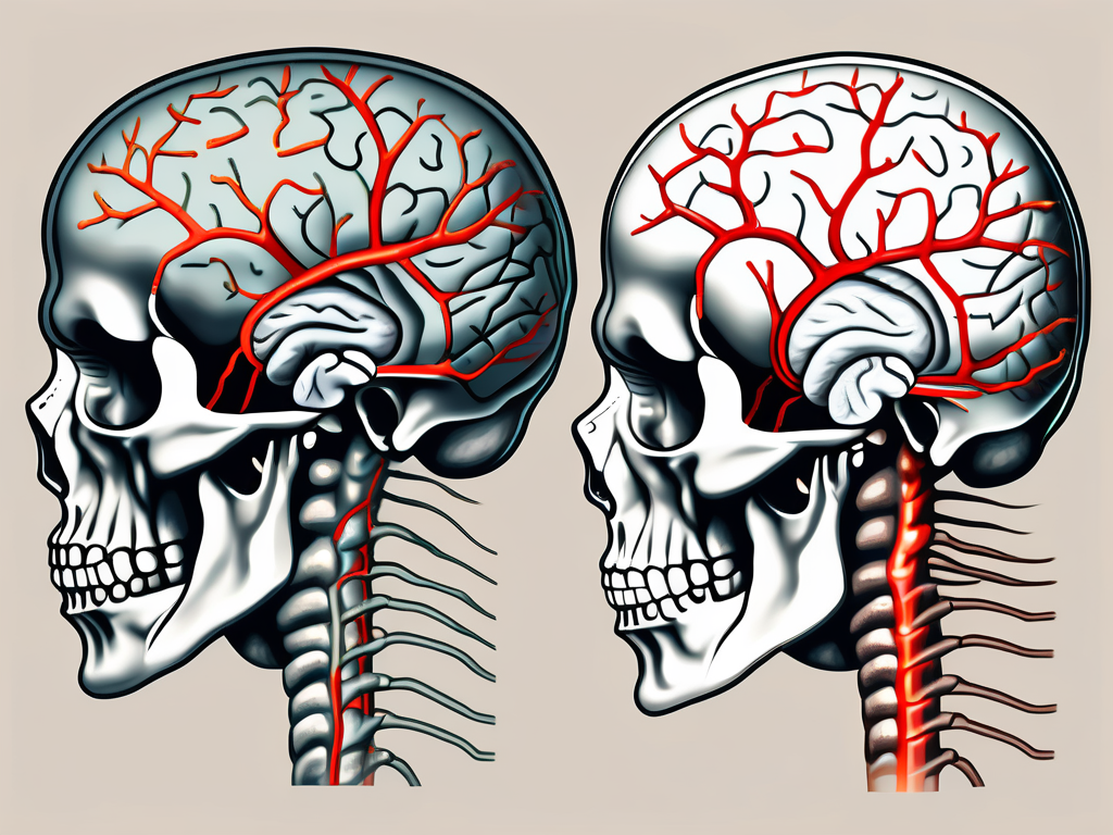
The trochlear nerve, also known as the fourth cranial nerve, is a critical component of our nervous system. It plays a crucial role in controlling the movement of one of our extraocular muscles, the superior oblique muscle, which enables the eye to rotate inwards and downwards. Understanding the anatomy, function, and clinical significance of the trochlear nerve is essential for medical professionals and individuals curious about their health. In this article, we will delve into the intricate details of the trochlear nerve, its pathway, clinical implications, and preventive measures for maintaining its well-being.
Understanding the Trochlear Nerve
The trochlear nerve is one of the twelve cranial nerves that play a crucial role in the functioning of the human body. It is responsible for controlling the movements of the superior oblique muscle, which allows for downward and inward eye movements. To fully comprehend the significance of the trochlear nerve, it is essential to explore its anatomy and understand its function.
Anatomy of the Trochlear Nerve
The trochlear nerve originates from the dorsal aspect of the midbrain, specifically the trochlear nucleus. This nucleus serves as the starting point for the nerve’s journey. As the nerve emerges from the posterior aspect of the brainstem, it takes a unique path by coursing dorsally around the cerebral peduncle. It is fascinating to note that the trochlear nerve is accompanied by the oculomotor nerve, another crucial cranial nerve responsible for controlling various eye muscles.
Continuing its journey towards the superior oblique muscle in the orbit, the trochlear nerve crosses obliquely along the brainstem. This distinctive trajectory sets it apart from other cranial nerves, making it easily identifiable. The intricate network of nerves and their pathways within the human body never ceases to amaze researchers and medical professionals alike.
Function of the Trochlear Nerve
The primary function of the trochlear nerve is to control the superior oblique muscle, which plays a vital role in eye movements. This muscle allows for downward and inward eye movements, contributing to the coordination and alignment of both eyes. When the trochlear nerve becomes impaired, whether due to injury or disease, it can result in weakened eye movements and consequently affect visual coordination.
It is important to note that the trochlear nerve primarily innervates one muscle, the superior oblique muscle. This unique characteristic makes it vulnerable to damage and can lead to distinct visual symptoms. Understanding the intricate relationship between the trochlear nerve and the superior oblique muscle is crucial for diagnosing and treating any issues that may arise.
Research and ongoing studies continue to shed light on the complexities of the trochlear nerve and its role in maintaining optimal eye function. The more we delve into the intricacies of the human body, the more we realize the remarkable nature of its design.
Pathway of the Trochlear Nerve
Origin and Course of the Trochlear Nerve
The trochlear nerve, also known as the fourth cranial nerve, plays a crucial role in eye movement. It originates from the trochlear nucleus, which is situated in the midbrain. This nucleus is located in the dorsal region of the brainstem, specifically in the tegmentum. The trochlear nucleus is unique among cranial nerve nuclei as it is the only one that is located on the contralateral side of the brainstem.
Exiting the brainstem dorsally, the trochlear nerve takes a distinctive pathway. It wraps around the cerebral peduncle, which is a bundle of nerve fibers that connects the midbrain to the rest of the brain. This looping course around the cerebral peduncle allows the trochlear nerve to reach its target muscle, the superior oblique muscle, which is responsible for downward and inward eye movement.
The lengthy pathway and particular course of the trochlear nerve set it apart from other cranial nerves. Its unique trajectory allows for precise control of eye movements, ensuring coordinated and accurate visual tracking.
Areas of the Brain Involved
While the trochlear nerve’s primary function is to control the superior oblique muscle, its impact extends beyond the midbrain. It interacts with various areas of the brain associated with visual processing, contributing to the integration of eye movements and visual perception.
One important structure that the trochlear nerve interacts with is the superior colliculus. The superior colliculus is located in the midbrain and plays a crucial role in visual attention and eye movement control. It receives input from the retina and other visual areas of the brain, including the occipital cortex.
The occipital cortex, located in the posterior region of the brain, is responsible for processing visual information. It is involved in tasks such as object recognition, color perception, and visual memory. The trochlear nerve’s connections with the occipital cortex highlight its role in maintaining optimal vision.
Overall, the trochlear nerve’s pathway and interactions with various brain regions demonstrate its importance in coordinating eye movements and visual perception. Without this cranial nerve, our ability to track objects, shift our gaze, and perceive the world around us would be compromised.
Clinical Significance of the Trochlear Nerve
The trochlear nerve, also known as the fourth cranial nerve, plays a crucial role in the movement of the eye. It is responsible for innervating the superior oblique muscle, which helps control the rotation and downward movement of the eye. Due to its unique anatomy and function, the trochlear nerve is susceptible to specific disorders that can significantly impact vision and ocular movement.
Common Disorders Affecting the Trochlear Nerve
One common disorder that can affect the trochlear nerve is trochlear nerve palsy. This condition occurs when the nerve becomes damaged or compressed, leading to a range of symptoms. Trochlear nerve palsy often manifests as a limitation in the movement of the affected eye, causing diplopia (double vision) and difficulty in maintaining appropriate visual alignment.
There are several potential causes of trochlear nerve palsy. Trauma, such as a head injury or orbital fracture, can damage the nerve and disrupt its normal function. Tumors, both benign and malignant, can also exert pressure on the trochlear nerve, leading to palsy. Additionally, vascular abnormalities, such as an aneurysm or arteriovenous malformation, can affect the blood supply to the nerve, resulting in its dysfunction.
While these disorders can be distressing, it is essential to consult with a healthcare professional for a comprehensive evaluation and accurate diagnosis. Prompt medical attention can help determine the underlying cause of trochlear nerve palsy and guide appropriate treatment options.
Symptoms of Trochlear Nerve Damage
Trochlear nerve damage often presents with specific symptoms related to ocular movement. One of the hallmark signs is vertical or torsional diplopia, which refers to double vision experienced in the vertical or rotational axis. This occurs because the affected eye is unable to properly align with the other eye, leading to overlapping images.
Compensatory head tilting is another common symptom of trochlear nerve damage. Individuals with trochlear nerve palsy may instinctively tilt their heads to alleviate the double vision. By tilting the head, they can align the eyes in a way that reduces the overlap of images, providing temporary relief.
In addition to diplopia and head tilting, trochlear nerve damage can cause headaches and eyestrain. The increased effort required to coordinate eye movements can lead to discomfort and fatigue, resulting in headaches and a feeling of eye strain.
If you are experiencing any of these symptoms, it is crucial to seek medical advice to determine the underlying cause and appropriate treatment. An ophthalmologist or a neurologist specializing in ocular disorders can conduct a thorough evaluation and recommend the most suitable management plan for trochlear nerve damage.
Diagnosis and Treatment of Trochlear Nerve Disorders
Diagnostic Techniques for Trochlear Nerve Disorders
In order to diagnose trochlear nerve disorders accurately, healthcare professionals may employ various diagnostic techniques. These can include a detailed medical history, comprehensive eye examination, specialized imaging, such as magnetic resonance imaging (MRI), and electrophysiological studies to assess the functioning of the nerve and associated structures.
During a detailed medical history, the healthcare professional will ask the patient about their symptoms, when they started, and any factors that may have triggered or worsened them. This information can provide valuable insights into the potential cause of the trochlear nerve disorder.
A comprehensive eye examination is another crucial diagnostic tool. It involves assessing visual acuity, eye movements, and coordination, as well as examining the structures of the eye. By carefully examining the eye, the healthcare professional can identify any abnormalities or signs of nerve damage.
In some cases, specialized imaging techniques, such as magnetic resonance imaging (MRI), may be necessary to visualize the trochlear nerve and surrounding structures in more detail. MRI uses powerful magnets and radio waves to create detailed images of the brain and nerves, allowing healthcare professionals to identify any structural abnormalities or lesions that may be affecting the trochlear nerve.
Electrophysiological studies can also be conducted to assess the functioning of the trochlear nerve and associated structures. These studies involve measuring the electrical activity of the nerve and muscles involved in eye movement. By analyzing the electrical signals, healthcare professionals can determine if there are any abnormalities or disruptions in the nerve’s functioning.
It is crucial to consult with a qualified healthcare practitioner for an accurate diagnosis and personalized treatment plan. They will carefully review the results of these diagnostic techniques and consider the patient’s individual circumstances to determine the most appropriate course of action.
Treatment Options for Trochlear Nerve Damage
The treatment approach for trochlear nerve damage depends on the underlying cause and severity of the condition. In some cases, conservative management strategies, such as eye patching or prism glasses, may be sufficient to alleviate symptoms and restore functionality.
Eye patching involves covering the unaffected eye to encourage the affected eye to work harder, which can help improve coordination and reduce double vision. Prism glasses, on the other hand, use specially designed lenses to redirect light and align images, reducing the strain on the trochlear nerve and improving vision.
However, more severe cases of trochlear nerve damage may require surgical intervention to correct or stabilize the affected eye muscle. Surgical procedures can involve repositioning or strengthening the muscle, or in some cases, releasing or adjusting the surrounding tissues to relieve pressure on the nerve.
Remember, each case is unique, and the appropriate treatment plan should be determined by a knowledgeable healthcare professional. They will consider factors such as the underlying cause, the severity of the symptoms, the patient’s overall health, and their individual preferences when developing a personalized treatment plan.
Prevention and Management of Trochlear Nerve Disorders
Lifestyle Changes for Trochlear Nerve Health
While trochlear nerve disorders can occur due to various factors beyond our control, certain lifestyle changes can promote overall ocular health. Maintaining a balanced diet rich in essential nutrients, engaging in regular exercise, and taking breaks from prolonged visual tasks can contribute to the well-being of the trochlear nerve and the entire visual system.
When it comes to maintaining a balanced diet, incorporating foods that are rich in omega-3 fatty acids, such as salmon, walnuts, and flaxseeds, can provide the necessary nutrients for optimal nerve function. Additionally, consuming fruits and vegetables that are high in antioxidants, like blueberries and spinach, can help protect the trochlear nerve from oxidative stress.
Regular exercise not only benefits overall health but also plays a crucial role in promoting trochlear nerve health. Engaging in activities such as walking, jogging, or swimming can improve blood circulation to the eyes, ensuring that the trochlear nerve receives an adequate supply of oxygen and nutrients.
Furthermore, taking breaks from prolonged visual tasks, such as staring at a computer screen or reading for an extended period, can help prevent strain on the trochlear nerve. Implementing the 20-20-20 rule, which involves looking away from the screen every 20 minutes and focusing on an object 20 feet away for 20 seconds, can significantly reduce eye fatigue and strain on the trochlear nerve.
However, always consult with a trusted healthcare professional for personalized advice tailored to your specific needs. They can provide guidance on the most suitable lifestyle changes to promote trochlear nerve health based on your individual circumstances.
Medical Interventions for Trochlear Nerve Disorders
In addition to lifestyle modifications, medical interventions may be necessary for the management of trochlear nerve disorders. These interventions aim to address the underlying cause of the condition and restore optimal ocular function.
One of the medical interventions commonly used for trochlear nerve disorders is medication. Depending on the specific condition, medications such as pain relievers, anti-inflammatory drugs, or muscle relaxants may be prescribed to alleviate symptoms and reduce inflammation or tension affecting the trochlear nerve.
Vision therapy is another medical intervention that can be beneficial for trochlear nerve disorders. This therapy involves a series of exercises and activities designed to improve eye coordination, strengthen eye muscles, and enhance visual processing. By targeting the underlying visual system issues, vision therapy can help alleviate symptoms associated with trochlear nerve disorders.
In severe cases where conservative treatments are ineffective, surgical procedures may be considered. Surgical interventions for trochlear nerve disorders aim to correct structural abnormalities or remove any obstructions that may be affecting the nerve’s function. These procedures are typically performed by experienced ophthalmologists or neurosurgeons who specialize in ocular and nerve-related conditions.
For an accurate diagnosis and appropriate treatment, it is vital to consult with an experienced healthcare provider. They will conduct a thorough evaluation of your symptoms, medical history, and perform any necessary diagnostic tests to determine the underlying cause of your trochlear nerve disorder. Based on their findings, they can recommend the most suitable medical interventions to manage and improve your condition.
Conclusion
Understanding and appreciating the significance of the trochlear nerve in our visual system is crucial. From its unique pathway and anatomical connections to its clinical implications and management, the trochlear nerve plays a vital role in maintaining optimal ocular health. If you suspect any issues with your eye movements or visual coordination, it is important to consult with a healthcare professional promptly. By doing so, you can gain proper diagnosis, access personalized treatment options, and embark on a journey towards preserving your trochlear nerve health.
