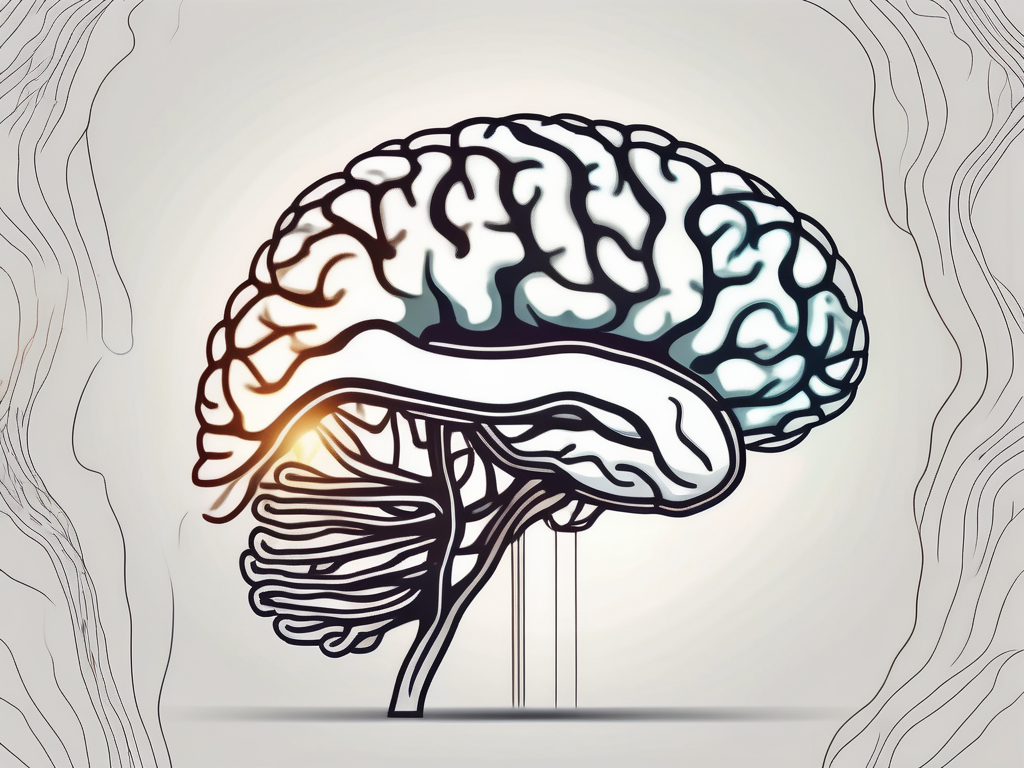
The trochlear nerve, one of the twelve cranial nerves in the human body, plays a crucial role in eye movement and coordination. Understanding the anatomy and function of this nerve is essential in order to appreciate its importance and the potential implications of any damage or disorders associated with it.
Understanding the Trochlear Nerve
Definition and Function of the Trochlear Nerve
The trochlear nerve, also known as the fourth cranial nerve or CN IV, is an essential component of the complex network that controls eye movement. It plays a crucial role in innervating the superior oblique muscle of the eye, which is responsible for various eye movements, including downward and outward eye movement, as well as rotation of the eye. This intricate coordination of eye movements ensures that our visual system functions optimally, allowing us to navigate the world around us with precision and accuracy.
When we think about the trochlear nerve, it is fascinating to consider its unique attachment point. Unlike most other cranial nerves that emerge from the ventral aspect of the brainstem, the trochlear nerve stands out by emerging from the posterior aspect. This distinctive anatomical arrangement adds to the intrigue and complexity of this nerve, making it a subject of great interest and study in the field of neuroanatomy.
The Unique Characteristics of the Trochlear Nerve
One of the most intriguing aspects of the trochlear nerve is its distinctive pathway around the brainstem. As it emerges from the posterior surface of the midbrain, it takes a fascinating route, looping around the brainstem before reaching its destination. This unique trajectory allows for precise control and coordination of eye movements, ensuring that our visual system functions harmoniously.
Furthermore, the trochlear nerve’s attachment point at the posterior aspect of the brainstem provides it with a strategic position to regulate eye movements. This unique anatomical arrangement allows the trochlear nerve to interact with other cranial nerves and brain structures involved in eye movement control, forming a complex network that ensures the smooth functioning of our visual system.
Studying the trochlear nerve and its intricate characteristics not only deepens our understanding of the human anatomy but also sheds light on the remarkable complexity and precision of the human visual system. By unraveling the mysteries of this nerve, researchers and medical professionals can gain valuable insights into various eye disorders and develop innovative treatments to improve visual health and quality of life.
Anatomy of the Trochlear Nerve
The trochlear nerve, also known as cranial nerve IV, plays a crucial role in eye movement and coordination. Let’s explore the origin, pathway, and its relationship with the eye muscles in more detail.
Origin and Pathway of the Trochlear Nerve
The trochlear nerve originates in the trochlear nucleus, a region located in the dorsal aspect of the midbrain. This nucleus is responsible for controlling the superior oblique muscle, one of the six extraocular muscles that control eye movement.
From the trochlear nucleus, the nerve fibers of the trochlear nerve cross the midline and decussate before exiting the brainstem. This unique decussation, or crossing over, contributes to the intricate pattern of innervation and eye movements associated with the trochlear nerve.
After exiting the brainstem, the trochlear nerve travels along the inner surface of the skull, protected by the meninges. It follows a complex pathway, navigating through bony canals and fibrous tunnels, until it reaches its final destination.
Eventually, the trochlear nerve reaches the superior oblique muscle, which it innervates. This muscle, located on the dorsal aspect of the eye, plays a crucial role in eye movement, particularly in downward and inward rotation.
The Trochlear Nerve and the Eye Muscles
Once the trochlear nerve reaches the eye muscles, it provides innervation specifically to the superior oblique muscle. This muscle is responsible for various eye movements, including depression, intorsion, and abduction.
The coordinated movement of the eyes is essential for visual perception and depth perception. The trochlear nerve works in conjunction with other cranial nerves, such as the oculomotor nerve (CN III), abducens nerve (CN VI), and optic nerve (CN II), to ensure smooth and precise eye movements in different directions.
Damage or dysfunction of the trochlear nerve can lead to a condition called trochlear nerve palsy, which can result in vertical or torsional diplopia (double vision) and difficulty in downward gaze. This condition requires careful evaluation and management by healthcare professionals specialized in ophthalmology and neurology.
In conclusion, the trochlear nerve is a vital component of the intricate network responsible for eye movement and coordination. Its origin in the trochlear nucleus, decussation, and innervation of the superior oblique muscle contribute to the complex interplay of cranial nerves that allow us to explore the world visually.
The Trochlear Nerve’s Connection to the Brain
The trochlear nerve, also known as cranial nerve IV, plays a crucial role in the control of eye movement. It is one of the twelve pairs of cranial nerves that emerge directly from the brain. The trochlear nerve has a distinctive attachment point in the brain, which allows for optimal control and coordination of eye movement.
As mentioned earlier, the trochlear nerve emerges from the posterior surface of the midbrain, specifically at the level of the inferior colliculus. This unique attachment point ensures that the nerve fibers do not need to travel extensively within the brain before reaching their destination. This efficient pathway allows for quick and precise communication between the trochlear nerve and the brain.
The midbrain, where the trochlear nerve originates, is a crucial region involved in the integration of sensory information and the coordination of motor responses. It plays a vital role in eye movement control, allowing us to track objects, shift our gaze, and maintain visual stability.
The Trochlear Nerve’s Attachment Point in the Brain
The posterior surface of the midbrain, where the trochlear nerve emerges, is a complex and intricate region of the brain. It is densely packed with various structures and pathways that contribute to its vital functions. One of the most prominent structures in this area is the inferior colliculus.
The inferior colliculus is a part of the auditory pathway, responsible for processing sound information. It receives inputs from the auditory nerve and plays a crucial role in sound localization and auditory reflexes. Interestingly, the trochlear nerve’s attachment point at the level of the inferior colliculus suggests a close relationship between auditory processing and eye movement control.
Studies have shown that the inferior colliculus not only processes auditory information but also receives visual inputs. This suggests that there might be a connection between the trochlear nerve and the visual processing centers in the brain. The precise nature of this connection and its functional implications are still subjects of ongoing research.
How the Trochlear Nerve Communicates with the Brain
The trochlear nerve communicates with the brain through various pathways and connections, ensuring the accurate control of eye movements and the maintenance of normal vision. Its communication network involves both efferent (outgoing) and afferent (incoming) signals.
When it comes to efferent signals, the trochlear nerve sends information from the brain to the muscles responsible for eye movement. These signals travel along the nerve fibers, originating from the trochlear nucleus in the midbrain. The trochlear nucleus is a small structure located in the midbrain, near the attachment point of the trochlear nerve. From there, the nerve fibers extend and innervate the superior oblique muscle of the eye, which plays a crucial role in eye movement control.
On the other hand, afferent signals refer to the feedback that the trochlear nerve receives from other cranial nerves and sensory receptors in the eyes. This feedback is essential for maintaining proper eye movement coordination and visual perception. The trochlear nerve integrates this feedback with the information it receives from the brain, ensuring smooth and accurate eye movements.
The intricate communication network involving the trochlear nerve, other cranial nerves, and sensory receptors in the eyes allows for the precise coordination of eye movements. This coordination is crucial for activities such as reading, driving, and playing sports, where accurate visual tracking and rapid shifts in gaze are essential.
Disorders Related to the Trochlear Nerve
The trochlear nerve, also known as the fourth cranial nerve, plays a crucial role in eye movement and coordination. When this nerve is damaged or dysfunctional, it can lead to various visual symptoms that can significantly impact an individual’s quality of life.
Symptoms of Trochlear Nerve Damage
One of the most common signs of trochlear nerve damage is double vision, also known as diplopia. This occurs particularly when looking downward or to the side. The affected individual may see two images instead of one, making it challenging to focus on objects effectively.
In addition to double vision, individuals with trochlear nerve damage may experience difficulty in moving the affected eye in specific directions. This can result in eye misalignment, making it challenging to coordinate eye movements and causing further visual disturbances.
Moreover, a reduced ability to focus on objects effectively, known as reduced visual acuity, is another symptom of trochlear nerve damage. This can make it difficult to read, drive, or perform other daily activities that require clear vision.
Diagnosis and Treatment of Trochlear Nerve Disorders
If you are experiencing any of the aforementioned symptoms, it is crucial to consult with a healthcare professional who specializes in neurology or ophthalmology. They will conduct a thorough examination to assess your eye movement and coordination, which will help guide the diagnosis.
In some cases, imaging techniques such as magnetic resonance imaging (MRI) may be utilized to assess the integrity of the trochlear nerve and associated structures. This can provide valuable information about the underlying cause of the nerve damage, such as trauma, inflammation, or compression.
Treatment options for trochlear nerve disorders vary depending on the underlying cause and severity of the symptoms. In some cases, medication may be prescribed to alleviate pain or reduce inflammation. Vision therapy, which involves exercises and techniques to improve eye coordination and movement, may also be recommended.
In severe cases where conservative treatments are ineffective, surgical interventions may be considered. These can include decompression of the trochlear nerve or repair of any structural abnormalities that are causing the nerve dysfunction.
It is important to note that only a qualified healthcare professional can determine the appropriate course of action based on your specific condition. Therefore, if you suspect trochlear nerve damage or are experiencing any related symptoms, seek medical attention promptly to receive an accurate diagnosis and appropriate treatment.
The Role of the Trochlear Nerve in Vision
How the Trochlear Nerve Affects Eye Movement
Eye movement is a complex process that involves several cranial nerves, including the trochlear nerve. The trochlear nerve specifically controls the superior oblique muscle, which aids in downward and outward eye movement. By contracting and relaxing this muscle, the trochlear nerve contributes significantly to the precise coordination and alignment of the eyes.
The trochlear nerve originates from the midbrain, specifically from the trochlear nucleus. It is the smallest cranial nerve and has the longest intracranial course. Exiting the brainstem dorsally, it wraps around the midbrain and enters the orbit through the superior orbital fissure. Once inside the orbit, the trochlear nerve innervates the superior oblique muscle, which is responsible for rotating the eye downward and outward.
Interestingly, the trochlear nerve has a unique attachment point in the brainstem. Unlike other cranial nerves that emerge from the ventral surface of the brainstem, the trochlear nerve emerges from the dorsal surface. This anatomical arrangement allows for the precise control and fine-tuning of eye movements.
The Impact of Trochlear Nerve Damage on Vision
When the trochlear nerve is damaged or malfunctioning, it can impact vision. The affected eye may not be able to move in specific directions, leading to a reduced field of vision and difficulty in performing various visual tasks. Trochlear nerve palsy, a condition characterized by the inability to move the affected eye downward and outward, is a common consequence of trochlear nerve damage.
Trochlear nerve damage can occur due to various factors, including trauma, infections, tumors, or even congenital abnormalities. In some cases, the damage may be temporary and resolve with time and appropriate treatment. However, in other cases, the damage may be permanent, requiring long-term management strategies to optimize visual function.
Diagnosing trochlear nerve damage involves a comprehensive evaluation of the patient’s medical history, a thorough physical examination, and specialized tests such as eye movement recordings and imaging studies. Treatment options depend on the underlying cause and severity of the nerve damage. In some cases, conservative management approaches, such as eye patching or prism glasses, may be sufficient to alleviate symptoms and improve visual function. However, more severe cases may require surgical intervention to correct the underlying issue or to reposition the affected eye muscle.
In conclusion, the trochlear nerve plays a vital role in eye movement and coordination. Its unique attachment point in the brain, together with its intricate pathways and connections, ensures precise control of the superior oblique muscle and contributes to overall visual function. Any signs of trochlear nerve damage or dysfunction should be promptly evaluated by a healthcare professional to ensure proper diagnosis and management. Remember, seeking expert medical advice is always the best course of action when faced with potential health concerns.
