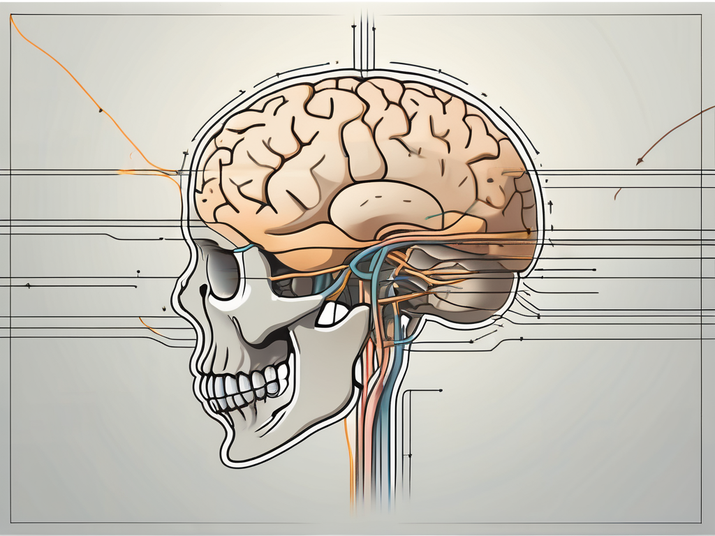
The trochlear nerve, also known as the fourth cranial nerve, is an important component of the human nervous system. Situated within the head, this nerve plays a crucial role in our vision and eye movement. In this article, we will delve into the complexities of the trochlear nerve, exploring its function, anatomical structure, associated disorders, and its contribution to our visual perception. By understanding the trochlear nerve, we can gain insight into the intricate workings of our visual system.
Understanding the Trochlear Nerve
Definition and Function of the Trochlear Nerve
The trochlear nerve is one of the twelve cranial nerves responsible for connecting the brain to different parts of the body. Specifically, it is the smallest cranial nerve and the only one that exits the brainstem from its dorsal surface. Like other cranial nerves, the trochlear nerve serves a distinct function. It innervates the superior oblique muscle of the eye, which aids in the downward and inward rotation of our eyes.
By controlling the eye movement, the trochlear nerve contributes to our ability to focus on objects at different distances and smoothly track moving targets. This intricate coordination is essential for our daily activities, facilitating tasks such as reading, driving, and even following a conversation.
Imagine sitting in a cozy coffee shop, sipping your favorite latte, and engrossed in a captivating novel. As you read, your eyes effortlessly glide across the words on the page, thanks to the trochlear nerve working behind the scenes. It ensures that your eyes move in perfect harmony, allowing you to absorb every word without strain or discomfort.
Furthermore, when you’re driving, the trochlear nerve enables you to maintain a clear view of the road ahead. It ensures that your eyes smoothly adjust to the changing distances between your vehicle and other objects, allowing you to navigate safely and confidently.
Anatomy of the Trochlear Nerve
To fully comprehend the functions and properties of the trochlear nerve, it is crucial to explore its anatomical structure. Originating from the dorsal aspect of the midbrain, in close proximity to the cerebral aqueduct, the nerve possesses a unique decussation pattern. Unlike most nerves, the fibers of the trochlear nerve cross to the opposite side of the brainstem before leaving the skull through the superior orbital fissure.
From there, the nerve travels along a defined pathway, ultimately reaching the superior oblique muscle of the eye. This complex trajectory of the trochlear nerve ensures the precise coordination necessary for efficient eye movement. Any disruption or damage to this pathway can result in debilitating vision impairments and related issues.
Picture a delicate dance between the trochlear nerve and the superior oblique muscle. As you look up at the night sky, the trochlear nerve sends signals to the superior oblique muscle, allowing your eyes to gaze at the twinkling stars above. This intricate connection between nerve and muscle ensures that your eyes move smoothly and effortlessly, enhancing your stargazing experience.
Moreover, the trochlear nerve’s unique decussation pattern adds an intriguing aspect to its anatomy. As the nerve fibers cross to the opposite side of the brainstem, they create a fascinating interplay between the two hemispheres of the brain. This interhemispheric communication contributes to the overall coordination and synchronization of eye movements, allowing us to explore our visual environment with precision and accuracy.
The Pathway of the Trochlear Nerve
Origin and Termination of the Trochlear Nerve
The trochlear nerve, also known as the fourth cranial nerve, originates from the nuclei located in the midbrain, specifically in the area near the superior colliculus. These nuclei are responsible for controlling the neurons that innervate the superior oblique muscle, one of the extraocular muscles involved in eye movement. The fibers of the trochlear nerve exit the midbrain dorsally and decussate, crossing over to the contralateral side of the brainstem.
After the decussation, the trochlear nerve proceeds through a long intra-axial course within the brainstem before emerging from the skull through the superior orbital fissure. This narrow passageway allows the nerve to connect with its target, the superior oblique muscle, which plays a crucial role in the coordination of eye movements.
The superior oblique muscle is responsible for rotating the eye downward and outward, allowing for precise eye movements and depth perception. The trochlear nerve’s innervation of this muscle enables the fine control necessary for these complex visual functions.
Structures Encountered by the Trochlear Nerve
While navigating its intricate pathway, the trochlear nerve encounters and interacts with various structures within the brain and skull. One such structure is the tentorium cerebelli, a thick, crescent-shaped fold of dura mater that helps separate the occipital lobes of the brain from the cerebral hemispheres. The trochlear nerve passes beneath this protective layer, ensuring its safe passage as it continues its journey.
Additionally, the trochlear nerve traverses through the cavernous sinus, a venous structure situated on the sides of the sella turcica, which houses the pituitary gland. This sinus is a complex network of veins and arteries, serving as a vital conduit for blood flow to and from the brain. The trochlear nerve’s proximity to this intricate vascular system highlights the importance of maintaining its integrity to ensure proper nerve function.
Furthermore, as the trochlear nerve travels through the superior orbital fissure, it intersects with numerous blood vessels, nerves, and connective tissues associated with the orbit. This region is densely packed with vital structures involved in vision, including the optic nerve, oculomotor nerve, and the various muscles responsible for eye movement. The trochlear nerve’s close proximity to these structures emphasizes the intricate interplay between the nerve and its surroundings, highlighting the delicate nature of its function and the potential for disruption when faced with certain disorders or injuries.
Disorders Associated with the Trochlear Nerve
The trochlear nerve, also known as cranial nerve IV, is responsible for controlling the superior oblique muscle of the eye. This muscle plays a crucial role in eye movement, particularly in looking downward and inward. However, like any other cranial nerve, the trochlear nerve is susceptible to damage or dysfunction, leading to various disorders.
Symptoms of Trochlear Nerve Damage
When the trochlear nerve is damaged, individuals may experience a range of symptoms that can significantly impact their daily lives. One of the most common symptoms is diplopia, also known as double vision. This double vision is often more pronounced when looking downward or inward, as these movements require the involvement of the superior oblique muscle.
In addition to diplopia, individuals with trochlear nerve damage may encounter difficulties in reading, navigating stairs, or performing tasks that require precise eye movements. The coordination between the eyes may be affected, leading to diminished depth perception. This can make judging distances and perceiving the three-dimensional world challenging.
Another symptom associated with trochlear nerve damage is eye misalignment. The affected eye may deviate from its normal position, causing a noticeable imbalance. To compensate for this misalignment, individuals may adopt abnormal head positions, tilting or turning their heads to align their eyes properly. These compensatory movements can be uncomfortable and may lead to neck or shoulder strain.
Diagnosis and Treatment of Trochlear Nerve Disorders
If you experience symptoms suggestive of trochlear nerve dysfunction, it is crucial to consult with a knowledgeable medical professional, such as a neurologist or ophthalmologist. These specialists have the expertise to conduct comprehensive evaluations and provide appropriate diagnosis and treatment.
During the evaluation, the medical professional will typically start by taking a thorough medical history to understand the onset and progression of symptoms. They will then perform a physical examination, which may involve assessing eye movements, checking for eye misalignment, and evaluating overall visual function. In some cases, additional tests such as neuroimaging studies may be ordered to assess the underlying cause of the trochlear nerve disorder.
Treatment options for trochlear nerve disorders vary depending on the underlying cause and severity of symptoms. In some cases, conservative measures may be prescribed to improve symptoms. Vision therapy, which involves exercises and activities aimed at improving eye coordination and control, may be recommended. Prism glasses, which can help align the images seen by both eyes and reduce diplopia, may also be prescribed.
In more severe cases, surgical intervention may be necessary. The specific surgical procedure will depend on the cause of the trochlear nerve damage. For example, if the misalignment of the eyes is the primary issue, surgery may be performed to correct the alignment and restore normal eye function. If the nerve damage is due to an underlying condition, such as a tumor or aneurysm, the surgery will focus on addressing the cause and relieving pressure on the nerve.
In conclusion, disorders associated with the trochlear nerve can cause a range of symptoms, including diplopia, difficulties in reading and performing precise eye movements, eye misalignment, and abnormal head positioning. Seeking medical attention from specialists in neurology or ophthalmology is essential for accurate diagnosis and appropriate treatment. With the right interventions, individuals with trochlear nerve disorders can improve their quality of life and regain visual function.
The Role of the Trochlear Nerve in Vision
The Trochlear Nerve and Eye Movement
Our ability to move our eyes precisely is essential for effective vision. The trochlear nerve plays a significant role in controlling the superior oblique muscle, primarily responsible for downward and inward eye rotation. This rotation allows us to maintain visual focus on objects below our straight line of vision.
But what happens when we look up? The trochlear nerve also assists in upward eye rotation by working in conjunction with other muscles, such as the inferior oblique and superior rectus muscles. This coordinated effort ensures that our eyes can explore the entire visual field, from the ground to the sky.
As we navigate our environment, the trochlear nerve ensures that our eye movements are coordinated and smooth, facilitating tasks such as reading, driving, and even following a moving target. Imagine trying to read a book without the precise control provided by the trochlear nerve. The words would blur together, making it nearly impossible to comprehend the text.
Furthermore, the trochlear nerve helps us maintain balance and spatial awareness. By working in harmony with other cranial nerves, it ensures that our eye movements align with our body’s position in space. This coordination is crucial for activities like walking on uneven terrain or reaching for objects without looking directly at them.
How the Trochlear Nerve Communicates with the Brain
The trochlear nerve maintains a unique communication pathway with the brain, ensuring continuous information flow. After the nerve fibers decussate at the level of the midbrain, they integrate into the existing neuronal circuits responsible for controlling eye movements. These circuits involve numerous other cranial nerves, such as the oculomotor and abducens nerves, which work in harmony to achieve coordinated eye movements.
But how does the trochlear nerve know where to send its signals? It receives input from various visual centers within the brain, such as the superior colliculus and the occipital lobe. These centers process visual information and send signals to the trochlear nerve, guiding its movements and ensuring that our eyes align with the objects we want to see.
By communicating with different centers within the brainstem and higher brain regions, the trochlear nerve helps regulate the complex interplay between visual stimuli and eye movement. This integration of sensory input and motor output ensures that our eye movements align with our visual perception.
Additionally, the trochlear nerve is involved in the pupillary light reflex. This reflex controls the size of our pupils in response to changes in light intensity. The trochlear nerve plays a role in coordinating the constriction of the pupil when exposed to bright light, protecting the delicate structures of the eye from potential damage.
In conclusion, the trochlear nerve is a vital component of our visual system, enabling precise eye movements and ensuring that our vision aligns with our perception of the world. Its intricate coordination with other cranial nerves and brain centers allows us to navigate our environment with ease and accuracy.
Frequently Asked Questions about the Trochlear Nerve
Common Misconceptions about the Trochlear Nerve
One common misconception regarding the trochlear nerve is that it is often overlooked when considering potential vision and eye movement issues. Due to its unique anatomical course and relatively uncommon involvement in isolated nerve damage, the trochlear nerve can be easily dismissed as a potential cause. However, it is essential for healthcare professionals to thoroughly evaluate this nerve when investigating such issues to provide accurate diagnoses and appropriate management.
When it comes to vision and eye movement, the trochlear nerve plays a crucial role. It is the fourth cranial nerve and is responsible for the innervation of the superior oblique muscle, which controls the downward and inward movement of the eye. Despite its importance, the trochlear nerve often flies under the radar, overshadowed by its more well-known counterparts like the optic nerve or the oculomotor nerve.
However, dismissing the trochlear nerve as insignificant can lead to misdiagnosis and ineffective treatment. In cases where patients present with symptoms such as double vision, difficulty looking downwards, or abnormal head tilting, healthcare professionals must consider the possibility of trochlear nerve involvement. By thoroughly evaluating this nerve, accurate diagnoses can be made, leading to appropriate management strategies and improved patient outcomes.
Future Research Directions for the Trochlear Nerve
The trochlear nerve continues to be an area of interest for researchers and medical professionals. Ongoing studies aim to elucidate the intricate details of its function, anatomical variability, and potential therapeutic approaches in cases of damage or dysfunction.
As our understanding of the trochlear nerve deepens, researchers are focusing on refining diagnostic techniques to better detect trochlear nerve-related issues. By developing more precise and sensitive tests, healthcare professionals can identify subtle abnormalities in the nerve’s function, leading to earlier intervention and improved patient outcomes.
Furthermore, future research may explore the efficacy of novel treatment modalities for trochlear nerve disorders. Currently, treatment options are limited, and there is a need for innovative approaches to address the specific challenges associated with trochlear nerve damage. By investigating new therapeutic strategies, such as targeted drug therapies or advanced surgical techniques, researchers hope to improve the prognosis for individuals affected by trochlear nerve-related conditions.
Additionally, understanding the interplay between the trochlear nerve and other components of the visual system is an area of interest for future research. The visual system is a complex network of structures and pathways that work together to facilitate vision and eye movement. Investigating how the trochlear nerve interacts with other cranial nerves, such as the oculomotor nerve or the abducens nerve, can provide valuable insights into the overall functioning of the visual system and potentially uncover new treatment approaches.
By expanding our knowledge in these areas, we can optimize patient care and improve outcomes for those affected by trochlear nerve-related disorders. The trochlear nerve may be often overlooked, but with continued research and attention, its significance in vision and eye movement can be fully appreciated, leading to better diagnosis, treatment, and overall patient care.
In Conclusion
The trochlear nerve, positioned within the complex network of cranial nerves, plays a crucial role in our visual system. Its involvement in eye movement coordination highlights its significance for our daily activities, ensuring seamless focus transitions and smooth tracking of moving objects.
Understanding the anatomy, function, and potential disorders associated with the trochlear nerve allows us to appreciate the intricacies of our visual perception. If faced with symptoms suggestive of trochlear nerve dysfunction, it is imperative to consult with a healthcare professional to receive appropriate evaluation and guidance.
Overall, the trochlear nerve is a remarkable component of our nervous system, contributing to our remarkable ability to explore and interpret the visual world around us.
