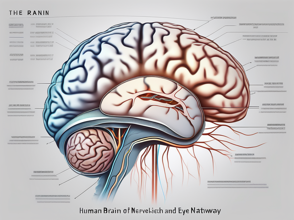
The trochlear nerve is a crucial component of the human nervous system. It plays a vital role in the control of eye movement and is responsible for innervating a specific muscle known as the superior oblique muscle. Understanding the intricate relationship between the trochlear nerve and this muscle is crucial for comprehending the complex mechanisms behind eye movement and for diagnosing and treating potential disorders related to this important neural pathway.
Understanding the Trochlear Nerve
Anatomy of the Trochlear Nerve
The trochlear nerve, also known as the fourth cranial nerve, is one of the 12 pairs of cranial nerves in the human body. It emerges from the dorsal surface of the midbrain, precisely from the trochlear nucleus.
The trochlear nucleus is a small group of neurons located in the midbrain, specifically in the tegmentum. It is positioned dorsally, near the cerebral aqueduct, which is a narrow channel that connects the third and fourth ventricles of the brain.
Unlike most other cranial nerves, the trochlear nerve has an atypical extracranial course, passing through a short space before entering the orbit via the superior orbital fissure.
The superior orbital fissure is a narrow opening located in the posterior part of the orbit. It serves as a passageway for several structures, including the trochlear nerve, as it makes its way from the midbrain to the eye.
Once in the orbit, the trochlear nerve follows a unique pathway around the posterior aspect of the eyeball. It then innervates the superior oblique muscle, allowing for precise control of eye movement, particularly in a vertical and rotational fashion.
The superior oblique muscle is one of the six extraocular muscles responsible for eye movement. It originates from the body of the sphenoid bone, near the optic canal, and inserts onto the sclera of the eye. Its primary function is to rotate the eye downward and inward.
Function of the Trochlear Nerve
The primary function of the trochlear nerve is to provide motor innervation to the superior oblique muscle. This muscle plays a vital role in the complex coordination of eye movements, allowing for optimal visual perception and stability.
By transmitting motor signals from the brainstem to the superior oblique muscle, the trochlear nerve enables the downward and inward rotation of the eyeball. This specific movement is essential for several visual tasks, including maintaining a stable focus when looking down or looking towards the nose, as well as coordinating binocular vision.
In addition to its motor function, the trochlear nerve also carries proprioceptive fibers. These fibers provide sensory information about the position and movement of the superior oblique muscle back to the brainstem, allowing for continuous feedback and adjustment of eye movements.
The trochlear nerve is susceptible to various pathological conditions that can affect its function. Lesions or damage to the nerve can result in a condition known as trochlear nerve palsy, characterized by weakness or paralysis of the superior oblique muscle. This can lead to double vision, difficulty in looking downward or inward, and an abnormal head tilt to compensate for the impaired eye movement.
In conclusion, the trochlear nerve is a crucial component of the visual system, providing motor innervation to the superior oblique muscle and playing a significant role in eye movement coordination. Its unique anatomical course and function highlight its importance in maintaining optimal visual perception and stability.
The Muscle Innervated by the Trochlear Nerve
Identifying the Superior Oblique Muscle
The superior oblique muscle is one of the six extraocular muscles responsible for controlling eye movements. It plays a crucial role in the intricate coordination of eye movements, allowing us to focus on objects and navigate our visual environment with precision.
Originating from the upper, anterior portion of the orbit, the superior oblique muscle follows a curved path through a fibrous ring known as the trochlea. This unique anatomical feature provides the muscle with a mechanical advantage, enhancing its ability to perform its functions effectively.
As the tendon of the superior oblique muscle loops through the trochlea, it gains leverage and stability, ensuring optimal control over eye movements. This arrangement allows the muscle to exert its rotational and depressant forces on the eye from the back, providing precise control over vertical eye movements.
Once the superior oblique muscle’s tendon passes through the trochlea, it inserts on the sclera – the tough, white outer layer of the eyeball. Specifically, it attaches at a point behind and slightly above the center of the eye. This strategic insertion point allows the muscle to exert its influence on the eye, facilitating coordinated movements and contributing to our ability to perceive the world around us accurately.
Role and Function of the Superior Oblique Muscle
The superior oblique muscle plays a pivotal role in the complex network of eye movements. Its primary function is to mediate the depression, rotation, and abduction of the eye. By working in harmony with the other extraocular muscles, it enables us to track moving objects, shift our gaze, and maintain visual stability.
When we look down, read, or engage in tasks that require close visual attention, the superior oblique muscle comes into action. It acts to rotate the eye inwardly and downwardly, allowing it to move towards the nose and downward in a coordinated manner. This precise control over eye movements is essential for tasks that demand fine motor skills and visual acuity.
Furthermore, the superior oblique muscle works synergistically with other extraocular muscles to maintain eye alignment and ensure precise binocular vision. This coordination is crucial for depth perception, as it allows the brain to fuse the images received from both eyes into a single, three-dimensional representation of the world.
In summary, the superior oblique muscle, innervated by the trochlear nerve, is a remarkable structure that contributes to the intricate system of eye movements. Its unique anatomical arrangement and precise control over vertical eye movements make it an indispensable component of our visual capabilities.
The Relationship Between the Trochlear Nerve and Superior Oblique Muscle
The trochlear nerve and the superior oblique muscle have a closely intertwined relationship that plays a crucial role in eye movement and visual function. Understanding how the trochlear nerve innervates the muscle and the implications of this connection is essential in comprehending the complexities of ocular motor control.
How the Trochlear Nerve Innervates the Muscle
The trochlear nerve, also known as the fourth cranial nerve, directly innervates the superior oblique muscle. It provides the necessary motor signals that control the movements of this muscle. The axons of the trochlear nerve exit the brainstem from the dorsal aspect and decussate, or cross over, to the opposite side just above the level of the brainstem.
Upon reaching the orbit, the trochlear nerve fibers enter the muscle cone, a fibrous structure that surrounds and protects the ocular muscles. Within the muscle cone, the trochlear nerve fibers navigate their way towards the superior oblique muscle, which is located on the upper, lateral aspect of the eyeball.
Once inside the superior oblique muscle, these nerve fibers distribute throughout its muscle fibers, forming a complex network of innervation. This intricate distribution allows for coordinated contractions necessary for precise eye movement control. The trochlear nerve ensures that the superior oblique muscle contracts with the appropriate force and timing to facilitate specific eye movements.
Implications of this Connection
The close anatomical and functional relationship between the trochlear nerve and the superior oblique muscle highlights the importance of this neural pathway in maintaining optimal eye movement and visual function. The trochlear nerve plays a crucial role in coordinating the movements of the superior oblique muscle, which is responsible for various eye movements, including depression, intorsion, and abduction.
Any disruption or damage to the trochlear nerve can lead to various ocular motor disorders and visual disturbances. Trochlear nerve palsy, for example, is a condition characterized by weakness or paralysis of the superior oblique muscle due to damage or dysfunction of the trochlear nerve. This can result in vertical diplopia (double vision), difficulty in looking downwards, and abnormal head positions to compensate for the impaired eye movement.
Understanding the relationship between the trochlear nerve and the superior oblique muscle is crucial in diagnosing and managing ocular motor disorders. Through careful examination and evaluation, healthcare professionals can identify and address any issues affecting this neural pathway, allowing for appropriate treatment and rehabilitation to restore optimal eye movement and visual function.
Disorders Related to the Trochlear Nerve
The trochlear nerve, also known as the fourth cranial nerve, plays a crucial role in eye movement. It is responsible for controlling the superior oblique muscle, which helps to move the eye downward and inward. When the trochlear nerve is damaged or dysfunctional, it can lead to a range of symptoms and conditions.
Symptoms of Trochlear Nerve Damage
Damage or dysfunction of the trochlear nerve can manifest in various ways, depending on the severity and extent of the injury or condition. Common symptoms of trochlear nerve damage may include:
- Diplopia (double vision): Individuals with trochlear nerve damage often experience double vision, particularly when looking downward or tilting their head. This double vision can be debilitating and significantly impact daily activities.
- Vertical misalignment of the eyes: Another common symptom is the misalignment of the eyes along the vertical axis. One eye may appear higher or lower than the other, leading to an asymmetrical appearance.
- Trouble looking down or inward: Due to the impaired function of the superior oblique muscle, individuals may find it challenging to look downward or inward. This can affect tasks such as reading, writing, and even walking down stairs.
- Headaches or eye strain: Trochlear nerve damage can cause frequent headaches and eye strain. The eyes may feel tired and overworked due to the extra effort required to compensate for the impaired eye movement.
If you experience any of these symptoms, it is crucial to consult with a medical professional, such as an ophthalmologist or neurologist, for a comprehensive evaluation and diagnosis. They can determine the underlying cause of your trochlear nerve damage and recommend appropriate treatment options.
Treatment and Management of Trochlear Nerve Disorders
The appropriate treatment approach for trochlear nerve disorders largely depends on the underlying cause and severity of the condition. In some cases, conservative management strategies such as patching the dominant eye or prism glasses may be recommended to alleviate the symptoms and improve visual alignment.
However, more severe cases may require surgical intervention to correct the alignment of the eyes and restore optimal eye movement control. Surgical options may include procedures to strengthen or reposition the superior oblique muscle or to address any other underlying structural issues.
It is essential to consult with a qualified healthcare professional to discuss the best treatment approach for your specific condition. They will consider factors such as your overall health, the severity of the trochlear nerve damage, and your individual needs and preferences.
Additionally, ongoing management and follow-up care are crucial for individuals with trochlear nerve disorders. Regular check-ups with your healthcare provider will allow for monitoring of your condition and adjustments to your treatment plan if necessary.
Furthermore, it is important to prioritize self-care and eye health. This can include practicing good eye hygiene, such as taking regular breaks from screen time, maintaining proper lighting conditions, and wearing appropriate eyewear when needed.
Living with trochlear nerve damage can be challenging, but with proper diagnosis, treatment, and management, individuals can experience improved quality of life and minimize the impact of their symptoms.
Prevention and Care for Optimal Nerve Health
Lifestyle Changes for Nerve Health
While specific prevention methods related to the trochlear nerve’s health are limited, maintaining overall nerve health is crucial for the optimal functioning of all neural pathways in the body. A healthy lifestyle that promotes general nerve well-being includes:
- Eating a nutritious diet rich in fruits, vegetables, and essential vitamins and minerals
- Engaging in regular physical exercise to improve circulation and overall nerve health
- Managing stress levels to minimize the impact of chronic stress on nerve function
- Maintaining a healthy weight to reduce the risk of nerve-related conditions such as diabetes
- Avoiding or minimizing the use of substances that can damage nerves, such as alcohol or recreational drugs
Adopting these healthy lifestyle habits may contribute to the overall well-being of the nervous system, including the trochlear nerve.
When it comes to maintaining optimal nerve health, it is important to consider the intricate network of nerves that exist throughout the body. Nerves play a vital role in transmitting signals between the brain and various parts of the body, allowing for essential functions such as movement, sensation, and coordination. By prioritizing nerve health, individuals can enhance their overall well-being and quality of life.
In addition to the lifestyle changes mentioned above, it is worth noting that certain nutrients are particularly beneficial for nerve health. For example, vitamin B12 is essential for the production of myelin, a protective sheath that surrounds nerve fibers and facilitates efficient signal transmission. Including food sources rich in vitamin B12, such as fish, meat, eggs, and dairy products, can help support nerve health.
Medical Interventions for Nerve Protection
In some cases, medical interventions may be necessary to protect and support the health of the trochlear nerve or other nerves in the body. These interventions may include:
- Regular check-ups with a healthcare professional to monitor nerve health and identify potential issues early
- Treating underlying medical conditions that can affect nerve function, such as diabetes or autoimmune disorders
- Consistently following prescribed medications and treatments for nerve-related conditions
It is important to note that discussing specific medical interventions and treatment options with a healthcare professional is crucial, as they can provide personalized advice based on your unique circumstances and medical history.
When it comes to nerve protection, early detection and intervention are key. Regular check-ups with a healthcare professional can help identify any potential issues before they progress and cause significant damage. By staying proactive and seeking medical guidance, individuals can take the necessary steps to protect their nerve health and overall well-being.
Furthermore, advancements in medical technology have led to various treatment options for nerve-related conditions. For instance, nerve stimulation techniques, such as transcutaneous electrical nerve stimulation (TENS), can help alleviate pain and promote nerve healing. These interventions are often tailored to the specific needs of the individual and may require the expertise of specialized healthcare professionals.
It is also important to recognize that nerve health is interconnected with overall body health. Conditions such as high blood pressure, heart disease, and certain autoimmune disorders can impact nerve function. By effectively managing these conditions through appropriate medical interventions and lifestyle modifications, individuals can support their nerve health and reduce the risk of complications.
Conclusion
The trochlear nerve’s connection to the superior oblique muscle is vital for the precise coordination of eye movements and overall visual function. Understanding the anatomy, function, and relationship between the trochlear nerve and the superior oblique muscle is essential for diagnosing and managing potential disorders related to this neural pathway.
If you are experiencing any symptoms or concerns related to eye movement or vision, it is crucial to seek guidance from a qualified healthcare professional. By working together with medical experts, you can ensure prompt evaluation, accurate diagnosis, and appropriate treatment options tailored to your specific needs, ultimately safeguarding optimal nerve health for a lifetime of visual well-being.
