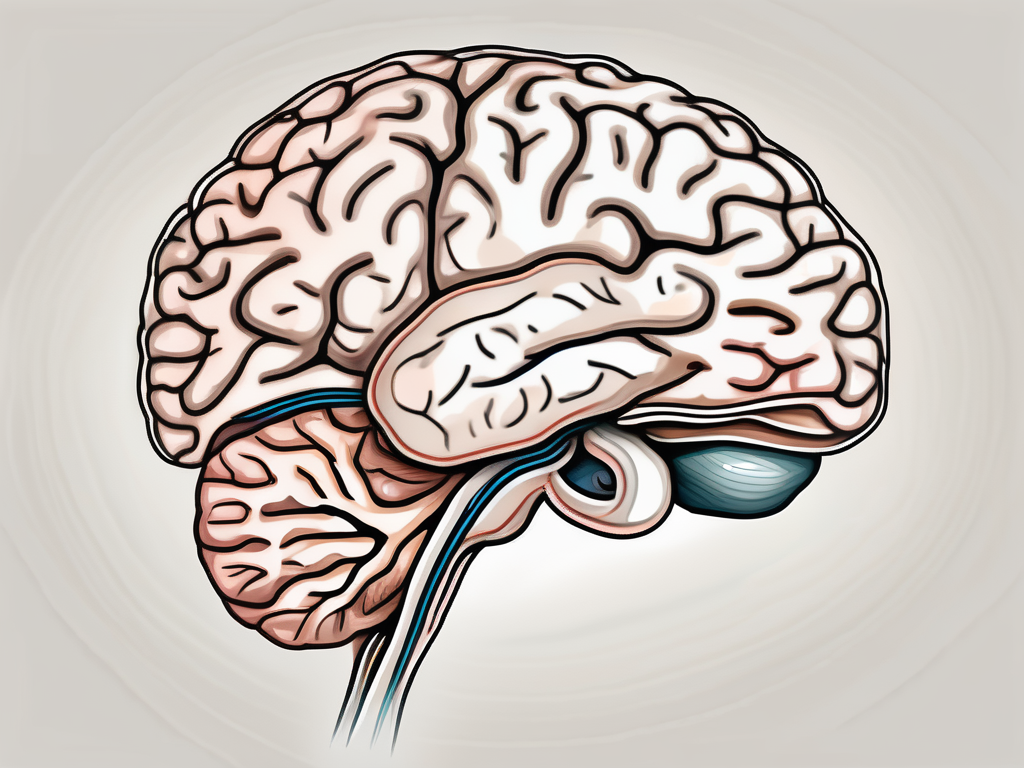
The trochlear nerve is one of the twelve cranial nerves responsible for the innervation of specific muscles in the human body. Understanding the role and function of this nerve is important in comprehending its significance in motor control and eye movement. Additionally, recognizing the disorders associated with the trochlear nerve and the corresponding diagnosis and treatment options can provide valuable insights into managing potential complications. In this article, we will explore the anatomy, function, associated disorders, and treatment modalities related to the trochlear nerve.
Understanding the Trochlear Nerve
The trochlear nerve, also known as cranial nerve IV, is a mixed nerve responsible for the innervation of the superior oblique muscle of the eye. It originates from the midbrain and runs posteriorly, traversing a unique pathway compared to other cranial nerves.
Emerging from the dorsal aspect of the brainstem, the trochlear nerve crosses over the tentorium cerebelli within the cavernous sinus, before reaching the superior oblique muscle. This anatomical arrangement allows for the precise control of eye movements.
The trochlear nerve is the smallest of all the cranial nerves and has the longest intracranial course. It is the only cranial nerve that emerges from the dorsal surface of the brainstem, making it vulnerable to certain types of brain injuries.
The trochlear nerve is unique in that it is the only cranial nerve to decussate, or cross over, within the brainstem. This decussation occurs at the level of the superior colliculus, which is responsible for integrating visual and auditory information. The decussation allows for the contralateral control of eye movements, meaning that the right trochlear nerve controls the left eye and vice versa.
Anatomy of the Trochlear Nerve
The trochlear nerve originates from the trochlear nucleus, which is located in the midbrain. It emerges from the dorsal aspect of the brainstem, just below the inferior colliculus. From there, it courses posteriorly and wraps around the cerebral peduncle before entering the cavernous sinus.
Within the cavernous sinus, the trochlear nerve runs alongside the oculomotor nerve (cranial nerve III), the abducens nerve (cranial nerve VI), and the ophthalmic and maxillary divisions of the trigeminal nerve (cranial nerve V). This close proximity to other important cranial nerves highlights the complex interplay between different structures involved in eye movements.
After traversing the cavernous sinus, the trochlear nerve exits the skull through the superior orbital fissure. It then enters the orbit and travels along the superior orbital wall, where it innervates the superior oblique muscle.
Function of the Trochlear Nerve
The primary function of the trochlear nerve is to enable downward and inward rotation of the eye. As the superior oblique muscle contracts, it acts synergistically with other extraocular muscles to facilitate coordinated eye movements.
The trochlear nerve plays a crucial role in rotational movements, allowing for the eyes to move in different directions. It is especially important for vertical gaze, as it controls the downward rotation of the eye. This is essential for activities such as reading, looking down at objects, or navigating uneven terrain.
In addition to vertical gaze, the trochlear nerve also contributes to convergence and divergence of the eyes during visual tracking of objects. This allows for the eyes to move together or independently, depending on the task at hand.
Damage or dysfunction of the trochlear nerve can lead to a condition known as trochlear nerve palsy. This can result in a variety of symptoms, including double vision, difficulty looking downward, and abnormal head posture to compensate for the impaired eye movements.
In conclusion, the trochlear nerve is a vital component of the intricate network of cranial nerves involved in eye movements. Its unique anatomical pathway and function allow for precise control of vertical gaze and rotational eye movements, contributing to our ability to perceive and interact with the world around us.
Muscles Innervated by the Trochlear Nerve
Superior Oblique Muscle
The trochlear nerve innervates the superior oblique muscle, which is one of the six extraocular muscles responsible for controlling eye movements. The superior oblique muscle originates from the apex of the orbit and inserts into the sclera near the posterior pole of the eye. It functions to depress, abduct, and internally rotate the eye. Dysfunction in the superior oblique muscle can lead to various visual disturbances and hinder the ability to effectively track moving objects.
The superior oblique muscle is a fascinating structure that plays a crucial role in the intricate dance of eye movements. Its unique origin and insertion points allow it to exert precise control over the eye’s position and orientation. When the superior oblique muscle contracts, it pulls the eye downward, away from the midline, and rotates it inward. This coordinated movement is essential for maintaining proper alignment of the eyes and ensuring accurate visual tracking.
Imagine a scenario where you are watching a fast-paced tennis match. As the players dart across the court, your eyes need to smoothly track the ball’s movement. This is where the superior oblique muscle shines. By working in harmony with the other extraocular muscles, it helps your eyes follow the ball’s trajectory with precision and accuracy.
Role of Trochlear Nerve in Eye Movement
The trochlear nerve plays a pivotal role in the complex coordination of eye movement. By controlling the superior oblique muscle, it aids in the fine-tuning of visual tracking and stabilization, especially during downward and inward gaze. This precise control is essential for maintaining binocular vision and depth perception. Dysfunction or injury to the trochlear nerve can result in diplopia (double vision), vertical misalignment, and difficulties in performing daily activities that require accurate eye movements.
When you look down to read a book or focus on a nearby object, the trochlear nerve and the superior oblique muscle work together to ensure that your eyes move smoothly and precisely. This coordinated effort allows you to maintain a clear and single image of the object you are looking at. Without the trochlear nerve’s contribution, your eyes would struggle to align properly, leading to blurred or double vision.
It is fascinating to think about the intricate network of nerves and muscles that enable our eyes to move effortlessly. The trochlear nerve, with its specific role in controlling the superior oblique muscle, is a vital player in this complex system. Understanding the functions and interactions of these structures not only deepens our appreciation for the marvels of human anatomy but also sheds light on the potential challenges that can arise when this delicate balance is disrupted.
Disorders Related to the Trochlear Nerve
Trochlear Nerve Palsy
Trochlear nerve palsy refers to the damage or dysfunction of the trochlear nerve, leading to abnormalities in eye movement. The trochlear nerve, also known as the fourth cranial nerve, is responsible for controlling the superior oblique muscle, which helps in downward and inward eye movements. When the trochlear nerve is affected, it can result in a range of symptoms and visual disturbances.
There are various causes of trochlear nerve palsy, including congenital malformations, trauma, inflammation, tumors, or vascular incidents affecting the nerve. In some cases, the condition may be present at birth (congenital) or acquired later in life. Trauma, such as head injuries or fractures involving the orbit, can lead to damage to the trochlear nerve, resulting in palsy.
Patients with trochlear nerve palsy often experience difficulties in looking downwards or medially, leading to a tilted head posture to compensate for the visual disturbance. This compensatory head tilt helps align the eyes and improve binocular vision. However, it can also cause neck strain and discomfort over time.
Diagnosing trochlear nerve palsy involves a thorough evaluation of the patient’s medical history, physical examination, and specialized tests. An ophthalmologist or a neurologist may perform a detailed eye examination, assessing eye movements, alignment, and visual acuity. Imaging studies, such as magnetic resonance imaging (MRI), may be ordered to identify any structural abnormalities or lesions affecting the trochlear nerve.
Management of trochlear nerve palsy depends on the underlying cause and the severity of symptoms. In some cases, the condition may resolve spontaneously without any treatment. However, if the palsy is causing significant visual impairment or interfering with daily activities, interventions may be necessary. Treatment options may include prism glasses to correct double vision, eye patching, or surgical procedures to address the underlying cause or improve eye alignment.
Causes and Symptoms of Trochlear Nerve Disorders
Trochlear nerve disorders can arise from various causes, including trauma, congenital malformations, tumors, or infections affecting the nerve. Trauma to the head or orbit, such as a direct blow or fracture, can lead to damage to the trochlear nerve and result in its dysfunction.
Congenital malformations of the trochlear nerve can occur during fetal development, leading to abnormalities in its structure or function. These malformations may be isolated or associated with other congenital conditions affecting the cranial nerves or the eye muscles.
Tumors, both benign and malignant, can also affect the trochlear nerve. These tumors may originate from the nerve itself or adjacent structures, such as the brainstem or the cavernous sinus. The presence of a tumor can compress or infiltrate the trochlear nerve, causing its dysfunction and resulting in various visual disturbances.
Infections, such as meningitis or sinusitis, can also involve the trochlear nerve and lead to its inflammation or damage. These infections may be bacterial, viral, or fungal in nature and can affect the nerve directly or through secondary mechanisms, such as immune-mediated reactions.
Common symptoms of trochlear nerve disorders include diplopia (double vision), especially when looking downward or inward. The double vision occurs because the affected eye is unable to properly align with the other eye, resulting in overlapping images. Eye misalignment, known as strabismus, may also be present, with one eye deviating or turning in a different direction than the other. This misalignment can cause difficulties in focusing, depth perception, and coordination of eye movements.
Reduced ability to perform tasks that require precise eye movements, such as reading or driving, is another common symptom. Patients may find it challenging to track moving objects or maintain a stable gaze. These visual disturbances can significantly impact daily activities and quality of life.
When experiencing symptoms suggestive of trochlear nerve disorders, it is crucial to seek medical attention for a thorough evaluation and establishment of the underlying cause. Early diagnosis and appropriate management are essential to minimize the impact on visual function and prevent further complications.
Diagnosis and Treatment of Trochlear Nerve Disorders
The trochlear nerve, also known as the fourth cranial nerve, is responsible for the innervation of the superior oblique muscle of the eye. This muscle plays a crucial role in eye movement, particularly in downward and inward rotation. When the trochlear nerve is affected by a disorder, it can lead to various symptoms such as double vision (diplopia), eye misalignment (strabismus), and difficulty in focusing.
Diagnostic Procedures
Diagnosing trochlear nerve disorders requires a thorough evaluation by a healthcare professional with expertise in neurology or ophthalmology. The process begins with a detailed medical history, where the healthcare provider will inquire about any previous eye injuries, surgeries, or underlying medical conditions that may contribute to the nerve dysfunction.
After the medical history, a comprehensive physical examination is conducted. This examination includes an assessment of eye movements, looking for any abnormalities or limitations in the range of motion. The healthcare provider may also perform specialized tests to further evaluate the function of the trochlear nerve.
Ocular motility assessment is one such test that involves tracking the movement of the eyes in different directions. This test helps identify any restrictions or weaknesses in the superior oblique muscle, which may indicate trochlear nerve involvement.
Visual field testing is another valuable tool in diagnosing trochlear nerve disorders. This test measures the extent and quality of a person’s peripheral vision. Any abnormalities in the visual field may suggest nerve dysfunction.
In some cases, neuroimaging studies, such as magnetic resonance imaging (MRI), may be ordered to visualize the structures of the brain and rule out any structural abnormalities or lesions that could be affecting the trochlear nerve.
These diagnostic procedures help healthcare professionals differentiate trochlear nerve involvement from other conditions that may present with similar symptoms, such as cranial nerve palsies or ocular muscle disorders.
Treatment Options and Rehabilitation
The management of trochlear nerve disorders depends on the underlying cause and the severity of symptoms. In some cases, conservative approaches may be sufficient to alleviate the symptoms and improve visual function.
Observation is often recommended in mild cases, where the symptoms are not significantly affecting daily activities. Regular follow-up appointments with the healthcare provider are necessary to monitor any changes in symptoms or progression of the disorder.
Prism glasses are another conservative treatment option for trochlear nerve disorders. These specialized glasses contain prisms that help correct the double vision caused by the misalignment of the eyes. By redirecting the light entering the eyes, prism glasses can help merge the two images into a single, clearer image.
Occlusion therapy is another approach used to manage diplopia. This therapy involves covering one eye with a patch or using special contact lenses to block the vision in the affected eye. By eliminating the input from one eye, the brain can focus on the image seen by the unaffected eye, reducing the perception of double vision.
In cases where conservative measures are inadequate or the symptoms are severe, surgical interventions may be necessary. Trochlear nerve decompression is a surgical procedure that aims to relieve any compression or entrapment of the trochlear nerve, allowing it to function properly. Strabismus surgery, on the other hand, is performed to correct the misalignment of the eyes and restore binocular vision.
Rehabilitation programs can also play a significant role in optimizing outcomes for individuals with trochlear nerve disorders. These programs often include visual exercises and eye muscle strengthening techniques to improve eye coordination and enhance visual function. Working with a specialized therapist or optometrist, individuals can regain control over their eye movements and reduce the impact of the disorder on their daily lives.
In conclusion, the trochlear nerve is a vital component of the complex neural network responsible for eye movement. Understanding the diagnosis and treatment options for trochlear nerve disorders is crucial in providing appropriate care and improving visual function. If you experience any symptoms related to the eyes or suspect trochlear nerve involvement, it is advisable to consult with a healthcare professional, such as an ophthalmologist or neurologist, who can provide an accurate diagnosis and guide you through the most suitable management strategies tailored to your specific needs.
