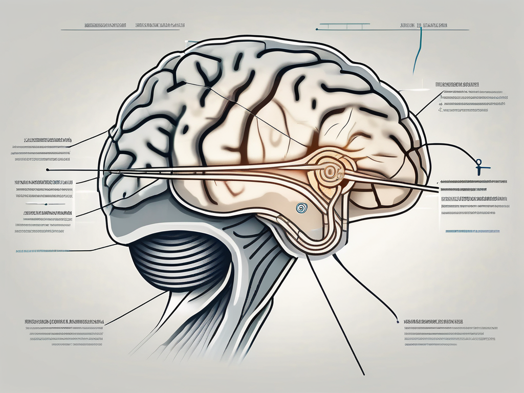
The trochlear nerve is a fascinating structure within the human body that plays a crucial role in our ability to move our eyes. Its name, however, might leave many individuals wondering about its origin and significance. In this article, we will delve into the intriguing world of the trochlear nerve, exploring its anatomy, function, and historical context. Join us on this enlightening journey to uncover why it is called the trochlear nerve.
Understanding the Trochlear Nerve
The trochlear nerve, also known as cranial nerve IV, is one of the twelve cranial nerves located within the brainstem. It is the smallest and longest cranial nerve, responsible for innervating the superior oblique muscle of the eye, which is crucial for eye movement and alignment. Emerging from the posterior aspect of the brainstem, the trochlear nerve takes a unique pathway, wrapping around the midbrain before reaching its destination.
As the trochlear nerve emerges from the brainstem, it traverses through a complex network of neural pathways, interacting with various structures along the way. These structures include the oculomotor nerve, which controls the movement of other eye muscles, and the trigeminal nerve, responsible for sensation in the face and motor functions such as chewing. The intricate interplay between these nerves ensures the precise coordination of eye movements and the maintenance of proper visual function.
Anatomy of the Trochlear Nerve
The trochlear nerve originates from the trochlear nucleus, a small group of neurons located in the midbrain. From there, it exits the brainstem dorsally, or towards the back, making it the only cranial nerve to do so. This unique pathway allows the trochlear nerve to avoid potential compression or damage from other structures in the brain.
As the trochlear nerve exits the brainstem, it wraps around the midbrain, forming a loop-like structure known as the superior medullary velum. This looping pathway provides the trochlear nerve with the necessary length to reach its target muscle, the superior oblique. The superior oblique muscle, located within the orbit of the eye, plays a vital role in eye movement, specifically in rotating the eyeball downward and outward.
Function of the Trochlear Nerve
The trochlear nerve primarily controls the movement of the eye by enabling the superior oblique muscle to rotate the eyeball downward and outward. This coordinated movement allows us to track moving objects, navigate our environment, and maintain proper eye alignment. Without the trochlear nerve’s precise control, our vision would be compromised, leading to difficulties in perceiving depth and accurately focusing on objects.
In addition to its role in eye movement, the trochlear nerve also contributes to the regulation of eye position. It helps to keep the eyes properly aligned, preventing conditions such as strabismus, commonly known as crossed eyes. The trochlear nerve’s ability to fine-tune the position of the eyes ensures that both eyes work together seamlessly, providing us with binocular vision and depth perception.
Furthermore, the trochlear nerve is involved in the coordination of eye movements with head movements. This integration allows us to maintain a stable visual field, even when our head is in motion. The trochlear nerve works in conjunction with other cranial nerves, such as the vestibulocochlear nerve, which is responsible for balance and hearing, to ensure that our visual system remains stable and responsive in various situations.
The Origin of the Name ‘Trochlear’
Etymology and Historical Context
The name ‘trochlear’ stems from the Latin word ‘trochlea,’ meaning pulley. This naming convention reflects the distinctive manner in which the trochlear nerve’s pathway loops around the trochlear nucleus, resembling a pulley mechanism. The term was first used by anatomists who marveled at the nerve’s intricate arrangement and drew inspiration from mechanical devices such as pulleys to describe its structure.
Exploring the historical context further, we find that the naming of anatomical structures has a rich tradition. Anatomists throughout history have often turned to familiar objects or concepts to describe the intricate workings of the human body. This practice not only helps in understanding the structure and function of these anatomical features but also adds a layer of poetic beauty to the scientific language.
Latin, being the language of science and academia during the Renaissance, provided a common ground for anatomists to name various structures. By using Latin terms, anatomists ensured that their discoveries could be understood and communicated across different regions and cultures.
The Connection to Anatomy
The choice to name the nerve after a pulley mechanism was not arbitrary. The trochlear nerve’s path around the midbrain, looping through a fibrous pulley known as the superior oblique tendon, ensures proper control and alignment of the eye’s movements. This remarkable anatomical feature is highlighted by the naming convention, emphasizing the nerve’s importance in the intricate choreography of our ocular function.
When we delve into the intricacies of ocular anatomy, we discover the remarkable interplay between various structures that allow us to see the world around us. The trochlear nerve, with its unique pathway, plays a crucial role in this intricate dance of vision. By looping around the trochlear nucleus and passing through the superior oblique tendon, it ensures that our eyes can move smoothly and precisely.
The superior oblique muscle, controlled by the trochlear nerve, is responsible for rotating the eye downward and outward. This movement is essential for our ability to focus on objects at different distances and angles. Without the trochlear nerve’s precise control, our eyes would not be able to perform these complex movements with such accuracy.
By understanding the connection between the name ‘trochlear’ and its anatomical significance, we gain a deeper appreciation for the wonders of the human body. The intricate design and precise functioning of our ocular system are a testament to the complexity and beauty of nature’s creations.
The Trochlear Nerve in the Human Body
The trochlear nerve, also known as cranial nerve IV, is one of the twelve cranial nerves that emerge directly from the brain. It is the smallest cranial nerve and has a unique pathway compared to the others. Unlike the other cranial nerves that emerge from the brainstem, the trochlear nerve emerges from the dorsal aspect of the midbrain, making it the only cranial nerve to exit from the posterior side of the brain.
Role in Eye Movement
As mentioned earlier, the trochlear nerve is responsible for coordinating the downward and outward movement of the eye. This movement is particularly crucial for tasks such as reading, driving, and sports activities that require hand-eye coordination. The trochlear nerve’s precise control allows us to effortlessly shift our gaze and maintain visual stability, enhancing our overall quality of life.
When we read, for example, the trochlear nerve helps us smoothly track the words on a page, ensuring that our eyes move in a coordinated manner from one line to the next. Without the proper functioning of the trochlear nerve, our eye movements would be jerky and uncoordinated, making reading a challenging and frustrating task.
In sports activities, such as playing tennis or basketball, the trochlear nerve plays a crucial role in allowing us to accurately track the movement of the ball. It enables us to anticipate the trajectory of the ball, adjust our position, and time our movements to successfully catch or hit it. Without the trochlear nerve’s precise coordination, our hand-eye coordination would suffer, affecting our performance in various sports.
Disorders Associated with the Trochlear Nerve
While the trochlear nerve performs its duties flawlessly in most individuals, certain conditions can affect its function, leading to various neurological disorders. Trochlear nerve palsy, for instance, results from damage or compression of the nerve, causing weakness or paralysis of the superior oblique muscle. This condition can disrupt eye movements, resulting in double vision, difficulty focusing, and problems with depth perception.
Individuals with trochlear nerve palsy may experience difficulties in performing everyday tasks that require precise eye movements, such as reading, driving, or even walking on uneven surfaces. They may also have trouble judging distances accurately, making it challenging to navigate their surroundings safely.
It is important to note that trochlear nerve palsy can occur due to various factors, including trauma, tumors, infections, or congenital abnormalities. Prompt diagnosis and appropriate treatment are essential to manage the underlying cause and improve the individual’s quality of life.
If you experience any abnormal changes in your vision, such as double vision, difficulty focusing, or problems with eye movements, it is crucial to consult with a healthcare professional for proper evaluation and guidance. They can perform a thorough examination, including neurological tests, to determine the underlying cause and recommend the most suitable treatment options.
In conclusion, the trochlear nerve plays a vital role in coordinating eye movements, allowing us to perform daily activities that require precise hand-eye coordination. Disorders affecting the trochlear nerve can significantly impact an individual’s visual function and overall quality of life. Seeking medical attention for any abnormal changes in vision is crucial to ensure timely diagnosis and appropriate management.
The Importance of the Trochlear Nerve
Impact on Daily Life
The trochlear nerve’s vital role in facilitating eye movement and alignment cannot be overstated. Its optimal functioning allows us to navigate our surroundings, perform daily tasks, and indulge in activities that bring us joy. The seamless coordination of our eyes, made possible by the trochlear nerve, enriches our visual experiences and ensures effective communication with the world around us.
Imagine a world where the trochlear nerve did not exist. Simple tasks such as reading a book, driving a car, or even crossing the street would become incredibly challenging. Our eyes would struggle to move in sync, resulting in blurred vision and a constant feeling of disorientation. The trochlear nerve, with its intricate network of fibers, ensures that our eyes work together harmoniously, allowing us to perceive the world with clarity and precision.
Furthermore, the trochlear nerve plays a crucial role in depth perception. It enables us to accurately judge distances, making activities like catching a ball or pouring a glass of water effortless. Without the trochlear nerve’s precise control over eye movements, our ability to interact with the physical world would be severely compromised.
Trochlear Nerve in Medical Science
Medical science remains deeply intrigued by the trochlear nerve, continually exploring its intricate workings and potential therapeutic interventions for related disorders. Researchers are continually striving to deepen our understanding of the nerve’s physiology, seeking new treatment modalities and approaches to improve patients’ well-being.
One area of particular interest is the study of trochlear nerve palsy, a condition characterized by the inability to move the affected eye in certain directions. This condition can significantly impact a person’s quality of life, making simple tasks like reading, driving, or even watching television challenging. Medical professionals are dedicated to finding innovative ways to restore the function of the trochlear nerve in individuals with palsy, utilizing techniques such as surgical interventions, physical therapy, and even advancements in neuroprosthetics.
Moreover, the trochlear nerve’s involvement in various neurological disorders, such as migraine and strabismus, has sparked significant research efforts. Scientists are investigating the underlying mechanisms that contribute to these conditions, aiming to develop targeted therapies that can alleviate symptoms and improve patients’ overall well-being.
Additionally, advancements in imaging technology have allowed researchers to visualize the trochlear nerve in unprecedented detail. High-resolution imaging techniques, such as magnetic resonance imaging (MRI) and computed tomography (CT), provide valuable insights into the nerve’s structure and function. These advancements have paved the way for more accurate diagnoses and personalized treatment plans, ensuring that patients receive the most effective care possible.
In conclusion, the trochlear nerve’s importance extends far beyond its role in eye movement and alignment. It is an intricate component of our daily lives, enabling us to navigate the world with ease and engage in activities that bring us joy. In the realm of medical science, the trochlear nerve continues to captivate researchers, driving advancements in understanding and treatment. As our knowledge of this remarkable nerve deepens, so too does our ability to enhance the lives of those affected by trochlear nerve-related conditions.
Frequently Asked Questions about the Trochlear Nerve
The trochlear nerve, also known as the fourth cranial nerve, is a fascinating and complex structure that plays a crucial role in our visual system. It is responsible for the innervation of the superior oblique muscle, which controls the movement and alignment of the eye. In this section, we will address some common misconceptions about the trochlear nerve and explore future research directions in this field.
Common Misconceptions
There are some common misconceptions surrounding the trochlear nerve that deserve clarification. One such misconception is that trochlear nerve palsy is always a result of a traumatic injury. While trauma can indeed cause nerve damage, there are other causes, such as congenital malformation, inflammation, or underlying medical conditions. It is important to understand that trochlear nerve palsy can occur due to various factors, and proper evaluation by a healthcare professional is necessary to determine the underlying cause and guide appropriate treatment.
Furthermore, another misconception is that trochlear nerve palsy only affects eye movements. While it is true that the trochlear nerve primarily controls the superior oblique muscle, its impact goes beyond just eye movements. The trochlear nerve also plays a role in maintaining proper eye alignment, depth perception, and visual stability. Any disruption in the function of this nerve can lead to visual disturbances and affect overall visual perception.
Future Research Directions
The trochlear nerve continues to captivate the scientific community, inspiring researchers to explore new avenues in the study of ocular motility and neurology. With the advancement of technology, researchers are now able to delve deeper into the intricacies of this nerve and its role in visual processing. Future research aims to unravel the molecular mechanisms underlying trochlear nerve development, regeneration, and repair.
Additionally, researchers are investigating the potential therapeutic interventions for trochlear nerve injuries and disorders. Novel treatment approaches, such as gene therapy, stem cell transplantation, and neuroprotective agents, are being explored to restore trochlear nerve function and improve visual outcomes. These exciting research endeavors hold promise for individuals with trochlear nerve-related conditions, offering hope for better diagnostic tools and more effective treatment options.
In conclusion, the trochlear nerve’s name derives from its unique pulley-like pathway around the midbrain, illustrating its significant role in eye movements and alignment. This cranial nerve is essential for our daily visual experiences and plays a central part in our well-being. By deepening our understanding of the trochlear nerve and its function, we can appreciate its remarkable role in our lives and recognize the importance of seeking professional medical advice when faced with potential issues related to this vital structure.
